Universal Tissue Clearing Kit
About the Universal Tissue Clearing Kit
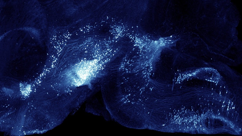
The Universal Tissue Clearing Kit is a reagent kit designed to enable reliable, reproducible, and high-quality tissue clearing across a wide range of biological samples. It is the ideal choice for next-generation 3D histology across a variety of tissues, including brain, spinal cord, liver, and lung. Built on a modified iDISCO-based protocol, it allows researchers to render intact tissues optically transparent. The protocol preserves proteins and their epitopes, which can be labeled using antibodies. The kit can also be used to look at the endogenous fluorescence of small molecule fluorophores, which are preserved.
The kit is easy to use and flexible, supporting the application of user-defined antibodies, requiring no specialized equipment, and providing a streamlined, reliable, step-by-step workflow that makes advanced 3D histology achievable in standard laboratory settings. By combining reproducible chemistry with clear protocol guidance, the kit lowers the technical barrier to whole-organ imaging while maintaining the quality required for quantitative, systems-level analysis.
The Universal Tissue Clearing Kit is the ideal choice for:
- Rapidly implementing tissue clearing in your own lab with a validated, easy-to-follow workflow
- Eliminating protocol guesswork with resources, such as a protocol calendar estimation
- Generating high-quality, reliable, and reproducible whole-organ 3D datasets

Looking for more information?
See Translucence and Bruker experts showcase the complete workflow in action.
Frequently Asked Questions
Tissue clearing uses a series of chemical steps to remove light-scattering lipids and match the sample’s refractive index. This process makes the tissue optically transparent, without disturbing its overall structure. When combined with antibody labeling and light sheet microscopy, you can image entire intact tissue. Using these methodologies allows scientist to preserve the spatial context of their samples and allows for unbiased, whole‑sample quantification.
The Universal Tissue Clearing Kit uses an organic solvent–based clearing method and is adapted from the iDISCO+ protocol. This approach produces excellent clearing across whole mouse brains and other tissues. The kit was created with Ricardo Azevedo, Translucence Biosystems CTO, who helped develop the iDISCO+ protocol.
Solvent‑based clearing quenches the fluorescence of most native fluorescent proteins. To visualize a fluorescent reporter (e.g. GFP, tdTomato), we recommend immunostaining with antibodies against your protein of interest. The kit’s buffers are fully compatible with immunolabeling, and signal amplification ensures robust detection of fluorescent proteins and other antigens.
The universal kit can clear and stain a wide range of tissues including mouse brains, rat brain hemispheres, spinal cords, lung, liver, kidney, organoids, and other soft tissues. With appropriate trimming and adaptations, the protocol can also be adapted for heart, eye and other organs. If you have questions about your specific tissue, contact our scientific support team (kits@translucencebio.com) for guidance.
The total protocol takes 26 days for a whole mouse brain. Much of this time is down time, and the total hands-on time is less than 3 working days. In the event that you sample of interest is smaller than a mouse hemisphere, the protocol can be considerably shortened. If you have a smaller sample that you would like to augment the protocol for, please reach out to kits@translucencebio.com for guidance.
The Universal Tissue Clearing Kit comes with enough reagents and tubes for clearing 5 samples. Clearing more than 5 samples? No problem! Solutions can be quickly added from individual kits to scale your reagents based on your experimental needs!
The Universal Tissue Clearing Kit Clearing requires only common laboratory equipment such as micropipettes, conical tubes, shaker or rotator for incubations, and a fume hood for handling organic solvents. You will also need access to a suitable microscope to image the cleared samples.
Samples should be perfused and fixed with paraformaldehyde using the same procedures you would follow for conventional IHC (e.g. transcardial perfusion with PBS followed by 4 % PFA). After perfusion, dissect and post‑fix the tissue overnight at 4 °C. Ensure that the tissue is thoroughly washed to remove excess fixative (3 x 1hr PBS washes). Store samples in PBS with .02% w/v sodium azide. Avoid freezing or using sucrose cryoprotection before clearing, as this will negatively impact the clearing process.
Yes. The kit includes pre‑made solutions, sample tubes, a detailed protocol, and supplementary materials like a protocol calendar that breaks the workflow into manageable steps (no weekend lab work required). Users without prior clearing experience, including students, have successfully cleared whole mouse brains using this protocol. Our goal is to provide a straightforward pathway to implementing tissue clearing without the need for extensive optimization.
There are several options for image analysis. Open‑source software such as ClearMap2 and BrainQuant3D can automatically detect labeled cells, register them to standard atlases and generate density maps from light‑sheet datasets. However, the implementation of these methods require a robust computational background and infrastructure. There are also Commercial packages like Imaris also offer 3‑D segmentation and quantification tools. If you prefer to outsource analysis, Translucence Biosystems offers AI‑powered 3D quantification services at discounted rates for their kit users. If you are interested in having Translucence Biosystems quantify your samples, reach out to kits@translucencebio.com.
Consistent perfusion-fixation, sample extraction, and imaging acquisition approaches are all essential for generating high-quality data. In the case of brains and other tissues that will be registered to a reference atlas, minimizing tissue damage is critical. Seemingly small cuts to the surface of a brain can drastically impact its ability to properly register. In terms of imaging, it is important to avoid introducing bubbles and to image the sample as close to the sagittal or horizontal orientation as possible.
The Translucence Biosystems support team is available to answer questions and provide troubleshooting advice throughout your tissue clearing journey. Email them at kits@translucencebio.com, and a scientist familiar with the protocol will respond promptly.
You can download the Universal Tissue Clearing Kit datasheet here.