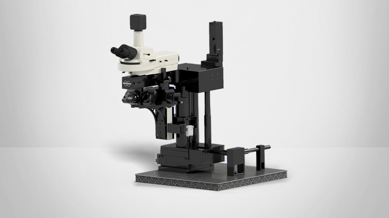
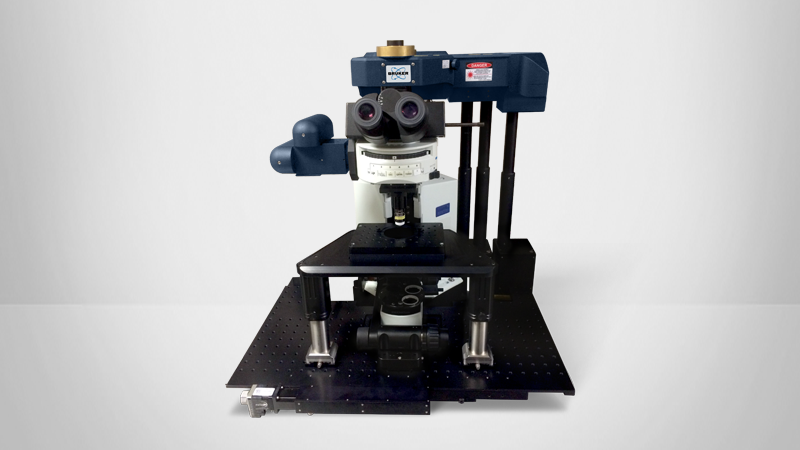
FLIM & PLIM Module
About Bruker's FLIM/PLIM Module
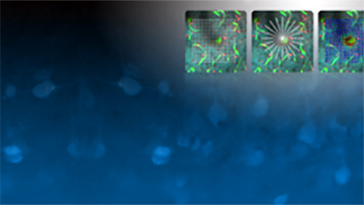
Many biological processes — including synaptic plasticity — involve dynamic interactions between proteins. Unfortunately, neither classical biochemical approaches nor normal diffraction-limited fluorescence microscopy provides the required sensitivity or resolution to study these intimate and sometimes transient interactions (1).
Contrasting these methods, Fluorescence Lifetime Imaging Microscopy (FLIM) delivers information about changes in the binding, conformation, and composition of biological samples that are not accessible using other techniques. It stands out as the most rigorous method for measuring:
- Molecular environment parameters;
- Protein-interactions by Fӧrster resonance energy transfer (FRET) (2); and
- The metabolic state of cells and tissue via their autofluorescence (3).


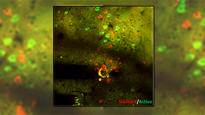
To do this, FLIM-based FRET measures the change in the decay function of the FRET donor on interaction with an acceptor. Similarly, Autofluorescence FLIM differentiates changes in the biology of objective cells and tissues based on the altered decay parameters of relevant endogenous fluorophores.
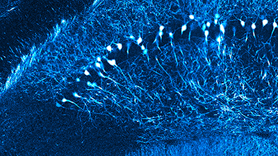
Likewise, Phosphorescence Lifetime Imaging Microscopy (PLIM) has become increasingly popular due to the recent development of two-photon-excitable oxygen-sensing probes (4). PLIM allows for quantitative in vivo oxygen measurements at high spatial and temporal resolution with high specificity, which makes it uniquely useful for live-cell and metabolic imaging.


How the FLIM/PLIM Module Supports Advanced Imaging
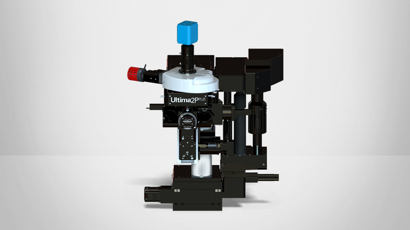
To leverage these unique capabilities, Bruker has developed a FLIM module based on Becker & Hickl 's FLIM systems. This module is fully integrated with all of Bruker’s multiphoton microscope systems, while Bruker’s proprietary Prairie View software enables advanced FLIM and PLIM data acquisition.
References
- 1. Datta R, Heaster TM, Sharick JT, Gillette AA, Skala MC. Fluorescence lifetime imaging microscopy: fundamentals and advances in instrumentation, analysis, and applications. J Biomed Opt. 2020 May;25(7):1-43. doi: 10.1117/1.JBO.25.7.071203.
- 2. Algar WR, Hildebrandt N, Vogel SS, Medintz IL. FRET as a biomolecular research tool - understanding its potential while avoiding pitfalls. Nat Methods. 2019 Sep;16(9):815-829. doi: 10.1038/s41592-019-0530-8.
- 3. Jones JD, Ramser HE, Woessner AE, Veves A, Quinn KP. Quantifying Age-Related Changes in Skin Wound Metabolism Using In Vivo Multiphoton Microscopy. Adv Wound Care (New Rochelle). 2020 Mar 1;9(3):90-102. doi: 10.1089/wound.2019.1030.
- 4. Esipova TV, Barrett MJP, Erlebach E, Masunov AE, Weber B, Vinogradov SA. Oxyphor 2P: A High-Performance Probe for Deep-Tissue Longitudinal Oxygen Imaging. Cell Metab. 2019 Mar 5;29(3):736-744.e7. doi: 10.1016/j.cmet.2018.12.022.