North American BioAFM User Meeting [2021]
AFM: An Important Tool for Biological Applications
FEATURING EXCLUSIVE PRESENTATIONS ON:
- Novel high-speed AFM techniques
- High-speed AFM fo studying cells in liquid
- AFM-based mechanobiology for cancer research
- AFM-based investigation of the structural properties of collagen
- AFM methods for characterizing structure-property relationships in oral biofilms
- In-vivo AFM for human skin disease diagnosis and monitoring in clinical settings
Featured Presentations & Technical Demonstrations
Simon Scheuring, Ph.D., Professor of Physiology and Biophysics in Anesthesiology, Weill Cornell Medicine
High-speed atomic force microscopy (HS-AFM) is a powerful technique that provides dynamic movies of biomolecules at work [1].
To break current temporal limitations to characterize molecular dynamics using HS-AFM, we developed HS-AFM height spectroscopy (HS-AFM-HS), a technique whereby we oscillate the HS-AFM tip at a fixed position and detect the motions of the molecules under the tip. This gives sub-nanometer spatial resolution combined with microseconds temporal resolution of molecular fluctuations. HS-AFM-HS can be used in conjunction with HSAFM imaging modes, thus giving access to a wide dynamic range [2]...
Heiko Haschke, Ph.D., Application Scientist, Bruker
Andreas Kraus, Ph.D., Application Scientist, Bruker
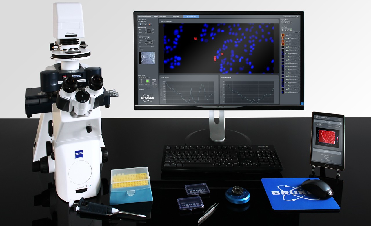
Recent atomic force microscopy (AFM) technology developments have led to unprecedented imaging rates in fluid, setting new milestones in high-speed scanning capabilities. Bruker recently launched the fastest commercially available high-speed AFM (NanoRacer®) able to reach a scanning speed of 50 frames/sec at 5000 lines/sec. High-speed AFM not only delivers atomic resolution but also enables the true, real-time visualization of time-resolved dynamics associated with cellular processes and the binding mechanisms of individual biomolecules. For example, the dynamics of individual protein binding behavior, two-dimensional protein assemblies, motor proteins, membrane trafficking, structural transitions of nucleic acids, can now be observed.
Kalpana Katti, Ph.D., University Distinguished Professor, North Dakota State University
The WHO reports about 3.4M cases of breast and prostate cancer each year, resulting in about a million deaths worldwide. The majority of the deaths are attributed to metastasis, the process of translocation, and recolonization of cancer to a remote site. Both prostate cancer and breast cancer exhibit a strong propensity to metastasize to bone, at which point the prognosis for patients is poor. Cancer, when diagnosed at the primary site, is treatable. However, more than 90% of cancer-related deaths result from metastasis. We have developed a novel bone mimetic nanoclay based scaffold to generate bone using the tissue engineering approach [1-5]. Seeding of commercial prostate and breast cancer cells on the tissue engineered bone is used to generate tumors [6, 7]. The tumors are investigated for mechanical properties using the direct nanoindentation technique...
.
Gunjan Agarwal, Ph.D., Professor, Mechanical and Aerospace Engineering, Ohio State University
In this study we investigated structural changes in collagen fibrils in human AAA tissue extracted at the time of vascular surgery and in aorta extracted from angiotensin II (AngII) infused ApoE−/− mouse model of AAA. Collagen fibril structure was examined using transmission electron microscopy and atomic force microscopy (AFM). Images were analyzed to characterize the length and depth of D-periodicity, fibril diameter and fibril curvature. Tissues were also stained using a novel reagent, collagen hybridizing peptide (CHP) (which stains degraded collagen) and analyzed using a combination of AFM and fluorescent microscopy. Our results elucidate how abnormal collagen fibrils with compromised D-periodic banding were observed in the excised human tissue and in remodeled regions of AAA in AngII infused mice...
Laurent Bozec, Ph.D., Professor (Associate), Faculty of Dentistry, University of Toronto
The oral cavity supports the growth of a diverse range of bacteria with its relatively stable environmental conditions and stream of nutrients derived from salivary components, gingival crevicular fluid and host diet. Oral biofilm formation on dental surfaces initiates with the attachment of primary colonizers followed by the attachment of secondary and later colonizers. Understanding how primary colonizers invade oral surfaces can be challenging but remain necessary for the development of new anti-microbial/anti-biofilm strategies. In this presentation, Dr. Bozec will review some of his team’s recent work on the biophysical characterization of bacterial species as well as the early-adhesions of oral bacteria/fungi on relevant surfaces characterized by single-cell force spectroscopy. In a second part of this presentation, Dr. Bozec will demonstrate how the combined used of Atomic Force Microscopy and Optical Coherence Tomography can be used pertinently to investigate the structure-property relationships in oral biofilms.
Mojtaba Azadi, Ph.D., Associate Professor, San Francisco State University
Detecting mechanical properties of the intact skin in-vivo leads to a novel quantitative method to diagnose skin diseases and to monitor skin conditions in clinical settings. Current research and clinical methods that detect skin mechanics have major limitations. The in-vitro experiments are done in non-physiological conditions and in-vivo clinical methods measure unwanted mechanics of underneath fat and muscle tissues but report the measurement as skin mechanics. An ideal skin mechanics should be captured at skin scale (i.e., micron-scale) and in-vivo. However, extreme challenges of capturing the in-vivo skin mechanics in micron-scale including skin motion due to heart beep, breathing and movement of the subject, has hindered measurement of skin mechanics in-vivo...


Speakers
Simon Scheuring, Ph.D., Professor of Physiology and Biophysics in Anesthesiology, Weill Cornell Medicine
Simon Scheuring is Professor of Physiology and Biophysics in Anesthesiology at Weill Cornell Medicine, New York, USA. He is a trained biologist from the Biozentrum at the University of Basel, Switzerland (1992−1996). During his Ph.D. (1997−2001) in the laboratory of Andreas Engel, he learned electron microscopy and atomic force microscopy and got interested in membrane proteins. During this period, he worked on the structure determination of aquaporins and sugar transporters. During his postdoc (2001−2004) and research assistant (2004−2007) at the institut national de la santé et de la recherche médicale (INSERM) and the Institut Curie in Paris, France, in the laboratory of Jean-Louis Rigaud, he learned membrane physical chemistry and developed atomic force microscopy for the study of native membranes. As junior research director (2007−2012), he set up his lab at the Institut Curie in Paris, France. Next, he built a larger independendent laboratory at INSERM/Aix-Marseille Université (2012) in Marseille, France, where he was promoted to senior research director (2012-2016). He then moved to Weill Cornell Medicine, New York, USA (2017), where he got appointed as Professor of Physiology and Biophysics in Anesthesiology.
Simon Scheuring has been awarded an INSERM Avenir (2005), the médaille de la Ville de Paris (2007), a European Research Council (ERC) consolidator grant (2012), the grand prix Robert Debré (2013), and the NIH director's pioneer award (2019). His objective is to head a dynamic research team with members from different scientific fields ranging from biology, physics, and engineering to develop and use atomic force microscopy-based technologies to provide novel insights into the processes taking place in biological membranes.
Kalpana Katti, Ph.D., University Distinguished Professor, North Dakota State University
Dr. Kalpana Katti is a University Distinguished Professor in the Department of Civil and Environmental Engineering at North Dakota State University. At NDSU, she has established a state-of-the-art materials characterization laboratory that houses advanced nanomechanical and infrared spectroscopic equipment as well as a Tissue Engineering Laboratory. She has served as PI on infrastructure development grants bringing advanced electron microscopes with federal funding that have led to significant expansion of the Electron Microscopy facility at NDSU. She also spearheaded a new doctoral program in Materials and Nanotechnology at NDSU. She leads the NDSU Center for Engineered Cancer TestBeds (CECT) and also the newly established NSF EPSCoR Center for cellular Biointerfaces in Science and Engineering. She has brought in research funding over $26M.
She has published 200 publications in Journals, conference proceedings and book chapters including 123 Journal articles. She has received the most cited award from the journal Colloids and Interfaces for the 2004-2007 years. She is also a NSF CAREER awardee. She is an elected Fellow of the American Institute for Medical and Biological Engineering (AIMBE). She received the Phi Kappa Phi lectureship in 2021, Waldron award for excellence in research in 2018, Peltier award for innovations in teaching in 2007, College of Engineering Researcher of the year 2003 and 2018, and the 52nd faculty lectureship award in 2011 at NDSU. She is also the recipient of the Roon award from Federation of Coatings Technologies in 1999 and Presidential Scholar award from Microscopy Society of America in 1996. She has given over 70 invited, keynote and plenary lectures at national and international conferences.
Gunjan Agarwal, Ph.D., Professor, Mechanical and Aerospace Engineering, Ohio State University
Dr. Gunjan Agarwal received her PhD in Biophysics from the Tata Institute of Fundamental Research in Mumbai, India and came to the US for her postdoctoral training at the Albert Einstein College of Medicine (Bronx, NY) followed by a year at Procter and Gamble Pharmaceuticals (Cincinnati OH). After a brief period as a research scientist at the Air Force Research Lab (WPAFB, OH), she joined the Ohio State University (Columbus, OH) as an Assistant Professor in 2003. Dr. Agarwal is currently a Professor of Mechanical and Aerospace Engineering at OSU with research focus in the domains of bioengineering and nanotechnology. She is an expert in the utilization of light, electron and atomic force microscopy (AFM) for biomedical research and directs a multi-user Bio-AFM core facility at OSU.
Her primary research focus is to study extracellular matrix remodeling with a particular emphasis of the collagen fibril structure and function. Another major initiative of Dr. Agarwal’s research program is to develop novel biomedical applications of the AFM. Her laboratory has applied many aspects of magnetic force microscopy (MFM) to study nanoparticle behavior and ferritin(iron) deposits in tissue sections in health and disease. She has coauthored over 50 journal articles and contributed four invited book chapters. Her research has been continuously funded by the National Institutes of Health, the National Science Foundation and the American Heart Association. She has mentored 10 Ph.D. and 5 MS students and teaches courses in Extracellular matrix, Medical Imaging and Microscopy.

Laurent Bozec, Ph.D., Professor (Associate), Faculty of Dentistry, University of Toronto
Professor Laurent Bozec received a BSc(Hons) in Applied Physics from the Nottingham Trent University (UK) in 1997, followed by a Ph.D. in Applied Physics and Instrumentation from Lancaster University(UK) in 2003. During his Ph.D. under the guidance of Professor Pollock, he developed a novel technique combining infrared spectroscopy and atomic force microscopy. Following his Ph.D., he joined both the University College London (UK) Faculty of Medicine and the newly formed London Centre for Nanotechnology. Under the guidance of the late Professor Mike Horton, he explored the nanoscale properties of collagen using Atomic Force Microscopy. In 2005, he published the first AFM image of collagen molecules using AFM.
In 2007, he joined the UCL Eastman Dental Institute where he started his own research group focusing on nanometrology of collagen and host-cell interactions. In 2012, he was one of the founding members of the Scanning Probe Microscopy section at the Royal Microscopical Society section in the UK. In 2015, he became the Vice-Dean Research at the UCL Eastman Dental Institute and created a doctoral school hosting at the time close to 100 Doctorate students. Over the years, Dr. Bozec has provided a high level of AFM expertise to both industrial clients and academic collaborators worldwide. Dr. Bozec has always been interested in Cultural Heritage and nanometrology. In 2014, he was fortunate to image one of Darwin’s notebooks leather book-binding with AFM and in 2017, he was able to image the skin of the 17th Cent mummified head of Henri IV of France. So far he has published over 120 manuscripts, totaling over 3500 citations.
In November 2018, Dr. Bozec relocated to the Faculty of Dentistry at the University of Toronto to create a translational research group focusing on the application of nanometrological tools to advance the knowledge of collagen-based connective tissue in health and diseases, such as Fibrosis, Cancer, Ehlers Danlos and natural aging.

Mojtaba Azadi, Ph.D., Associate Professor, San Francisco State University
Dr. Azadi holds a Ph.D. from the University of Alberta in Canada. He completed his master and undergraduate studies at Sharif University of Technology and Tehran universities in Iran.
He is an Associate Professor at the School of Engineering at San Francisco State University (SFSU) since August 2015. The primary goal of Dr. Azadi's lab is developing sensitive and quantitative biomechanical assessment tools to detect subtle changes in soft biological materials caused by biological factors, medications, and medical interventions.
Prior to joining SFSU, he worked for four years on numbers of collaborative projects around Biomimetics, Bioinstrumentation, Mechanobiology and Rehabilitation Robotics at the Biological and Mechanical Engineering Departments at MIT and Faculty of Rehabilitation Medicine at the University of Alberta. These projects include designing a Bio-tensegrity leg for a robotic cheetah, measuring mechanics of soft tissues such as skin, Fallopian tube, cartilage, Tectorial membrane, and different cell layers in micro and nano scales.
Andreas Kraus, Ph.D., Application Scientist, Bruker
Andreas Kraus is a physics graduate from the University of Leipzig, Germany, and received his Ph.D. from the Technical University of Braunschweig for his work on epitaxial growth and characterization of III-V semiconductor structures. In 2016 he joined JPK Instruments AG, as an applications scientist. Since 2019 he is a part of the worldwide Applications and Customer Support team at Bruker Nano, based in Berlin. He is currently focusing on high-resolution and high-speed applications of AFM for studying the dynamics of life sciences systems.

Heiko Haschke, Ph.D., Application Scientist, Bruker
Heiko Haschke received his Ph.D. in physics from the University of Bristol, UK, where he focused on the application and development of high-speed imaging and force spectroscopy for studying single biomolecular interactions. In 2001, he joined JPK Instrument AG as a developer of nanoanalytical instrumentation, in particular, AFMs and optical tweezers for biological applications. In 2004, he set up and became head of the Applications group, a team of applications scientist experts, providing worldwide customer and technical support. Since 2020, he is overseeing the worldwide applications development of the NanoRacer® high-speed AFM, from the BioAFM unit of Bruker Nano, based in Berlin, Germany.