HybridStage™ - Automated, Large Sample-Area Mapping Made Easy
Discover Our Versatile Solutions for Specialized Imaging
The mapping of tissue samples and thicker multi-cellular layers, as well as the nanomechanical characterization of the extracellular matrix and its embedded cells, requires a (semi)automated AFM approach for a millimeter range in x,y with nm–resolution. Large variations in the topography of tissue samples, combined with fluidity and stickiness of membrane and extracellular proteins, require greater flexibility in the z-axis of the AFM.
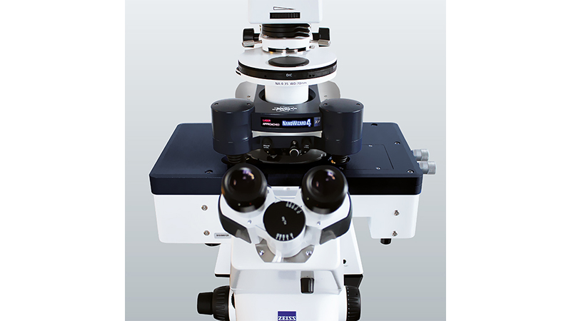
Our HybridStage—a modular, piezo-based sample scanner stage combined with motorized XY sample movement—provides a highly versatile solution to these challenges.
The HybridStage is a tool for advanced, multi-parametric AFM characterization of samples in the range of mm to nm and pN resolution, which features three scanner units that can move the tip/sample. A wide range of samples can be studied, including biomaterials, cells and macroaggregates, embryos and tissues, model organisms in developmental biology (zebrafish, C. elegans, etc), and implants. Particularly the increased z-piezo range, equipped with wide-ranged motorized sample motion, is perfect for the nanomechanical characterization of tissues required in cancer, nanomedicine, or developmental biology.
Readers can expect to learn about:
- The capabilities of the HybridStage;
- The advantages of large-range tiling of optical images; and
- How the HybridStage, together with NanoWizard software, facilitates multi-scan force mapping over large areas on very rough, very soft, and sticky samples.
KEYWORDS: Cell-Cell Interactions; Extracellular Matrix Proteins; Optical Tiling; Single Cell Force Spectroscopy; Tissue Mapping