For a better understanding of cardiovascular diseases
Cardiovascular diseases caused more than 19 million deaths globally in 20201
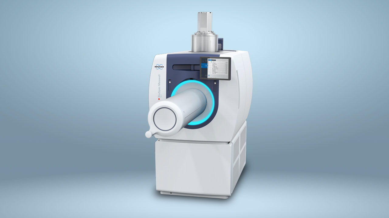
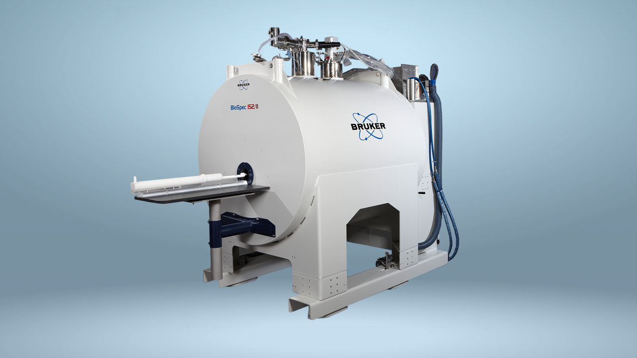
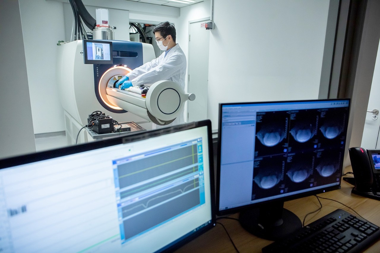
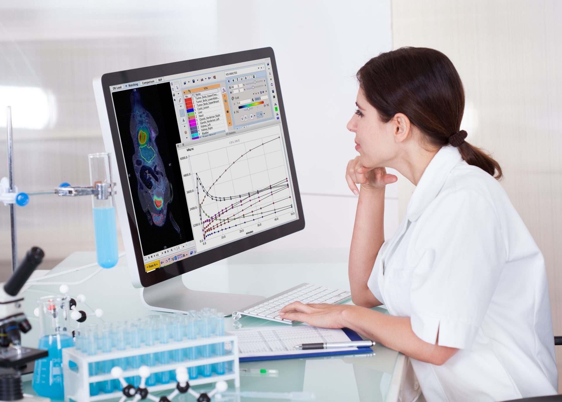
Preclinical cardiovascular research
Cardiovascular diseases (CVDs) caused approximately 19.1 million deaths globally in 20201 and, according to the World Health Organization (WHO), are the leading cause of death worldwide.2 This trend of high incidence and mortality is on the rise in low- to middle-income countries whose populations are living considerably longer than before and are therefore at higher risk of developing CVD.
Age is not the only risk factor, however. The underlying causes of different types of CVD, such as coronary heart disease and cerebrovascular disease, can be complicated and are not limited to diet and smoking. Poverty, stress, and environmental pollutants have been shown to influence risk of CVD.
Magnetic resonance imaging (MRI) is a widely used diagnostic method for CVD, and is also an important tool in preclinical research. By investigating animal models of CVD with magnetic resonance imaging techniques, such as magnetic resonance angiography, magnetic resonance spectroscopy (MRS), and magnetic resonance angiography (MRA), researchers gain a better understanding of disease pathophysiology and progression.
This non-invasive method allows longitudinal measurements over extended periods of time and provides a multitude of quantifiable parameters, such as ventricle volume or strain.
A new generation of MR technology
Preclinical MR is a well-established imaging technique to investigate a multitude of small animal cardiac disease models. While CT contrast agents (e.g. iodinated agents) typically used in clinical CT cardiograph protocols clear too quickly for use in small animal models, preclinical MR provides strong cardiac contrast for both enhanced and non-enhanced contrast imaging.
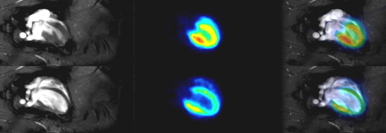
As mice and rats have heart rates up to 5x faster than humans, high-contrast CINE imaging with good slice and/or frame coverage is often required to resolve exquisitely small features of rodent cardiac tissue, and to obtain functional measurements such as ejection fraction.
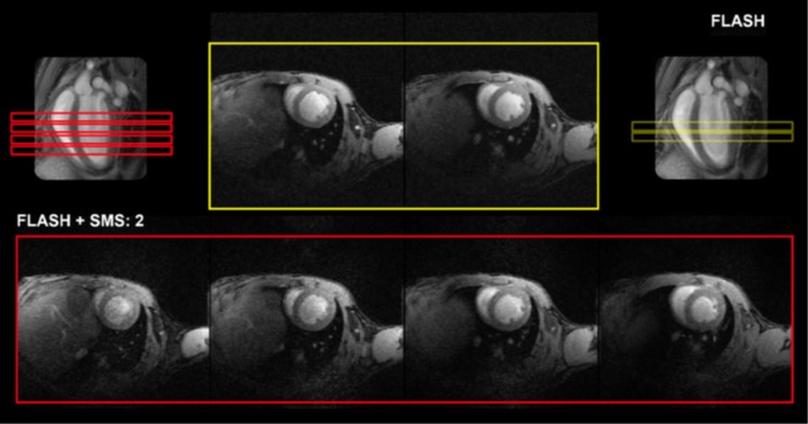
Accelerated SMS (Red) CINE imaging provides more slice coverage compared conventional (Yellow) CINE imaging without increasing scan time. Scans were acquired on a BioSpec 70/30 with a mouse cardiac array, and only varying SMS Factor (None or SMS2) and Slice Number (2 or 4). All other parameters were constant TE/TR: 1.6/10 ms, Res.: (130 x 130) μm2, Slice Thick.: 800 μm Movie Frames: 12, Acquisition Time (triggered): ~8 m 30 s.
Speeding up MR workflows
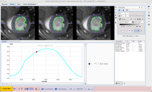
Preclinical MR hardware and software methods that allow for high signal-to-noise ratios (SNR) while minimizing total scan time can be highly beneficial in small animal cardiology. Bruker MR instrument hardware and methods, including ergonomically designed cardiac array coils, fast gradients and amplifiers, self-gated methods (i.e. IntraGate), and artificial intelligence (AI)-based segmentation, can reduce overall measurement times considerably, to increase cardiac imaging performance.
Beyond improvements of the preclinical cardiac measurement method itself, a streamlined and robust workflow from setup to evaluation of cardiac parameters makes cardiac investigations and analysis a straightforward process, supporting preclinical researchers.
References
- 2022 Heart Disease & Stroke Statistical Update Fact Sheet: Global Burden of Disease, American Heart Association. https://www.heart.org/-/media/PHD-Files-2/Science-News/2/2022-Heart-and-Stroke-Stat-Update/2022-Stat-Update-factsheet-GIobal-Burden-of-Disease.pdf [Accessed 11.02.22]
- Cardiovascular diseases (CVDs): Key facts. World Health Organization (WHO). June 2021. https://www.who.int/news-room/fact-sheets/detail/cardiovascular-diseases-(cvds) [Accessed 11.02.22]