Application Note: Single Molecule Localization Microscopy - Introduction & Applications
In this application note, readers will learn how Single Molecule Localization Microscopy (SMLM) surpasses the resolution limits of conventional light microscopes to deliver sharper, more detailed images of biological specimens. Discover how this super-resolution technique enables transformative insights across genomics, neuroscience, virology, and oncology through enhanced imaging and multiplexed labeling methods.
Contents include:
- Unlocking nanoscale insights with Single Molecule Localization Microscopy
- Using Bruker's Vutara super-resolution microscopy technology for advanced live and fixed imaging
- High-precision data capture and multicolor labeling with DNA-PAINT
- Application examples in genomics, neuroscience, virology, oncology, cardiology, and developmental and cell biology

Introduction
Since the early days of microscopic imaging, microscopes have been at the forefront of many biological discoveries and scientific advances, such as the first description of a cell1 and the discovery of bacteria,2 cholera, and anthrax.3 Since then, microscope technology has become increasingly powerful, allowing scientists to look at the world at a level previously unimaginable.
The light microscope is the most common microscope found in the laboratory; it works using visible light and a system of lenses to generate a magnified image of an object. However, light microscopes can be limited by low resolution; this can produce poor images and can cause intricate and potentially critical details of the specimen to be overlooked.4,5,6
Super-resolution techniques, such as single molecule localization (SML) microscopy, have been created to overcome some of the key challenges associated with light microscopy. These techniques can surpass the diffraction limit and generate higher quality images of biological specimens.

Applications
Super-resolution microscopy
There are three main types of super-resolution microscopy: structured illumination microscopy (SIM), stimulated emission depletion (STED) microscopy, and SML microscopy. SIM uses Moiré patterns and Fourier transforms to create a two-fold improvement on the resolution. STED reduces the size of the diffraction limited spot by using spread function engineering – this achieves a five- to ten-fold resolution improvement. SML offers the best improvement in resolution, along with quantitative and statistical analysis capabilities. SML microscopy takes a movie of an image, allowing information to be gathered from the image over time. The data from the SML allows spatial information to be abstracted and artificially generates an image below the diffraction limit, and it can be used in a wide array of scientific fields.5,7
Super-resolution microscopy is a very powerful tool for biological research. However, attaining multiplexed images for many distinct target species can sometimes be a challenge. SML microscopy can be combined with other techniques to overcome these obstacles.8 One such technique is DNA-PAINT (Point Accumulation in Nanoscale Topography). This approach enables single molecule localization through the binding of short (<10 nucleotides) fluorophore-labeled oligonucleotides to complementary oligonucleotides that are bound to a target molecule or nanobody. This binding creates a blinking effect, which produces high precision localization microscopy and multicolor imaging.9

Genomics
In genomics, SML can be used to study the functional organization of the genome. The organization of the genome within the nucleus can provide a lot of information on cellular pathology; in particular, it can help distinguish between pathological and non-pathological states. Understanding these differences can help facilitate research toward future medical diagnoses and treatment. However, work within this field has been hampered by the resolution limits of conventional light microscopy. By enabling the generation of highly detailed images using fluorescent methods,10,11 superresolution approaches such as SML microscopy have overcome these limitations.
Chromosomes are a collection of proteins and DNA that store genetic information. The three-dimensional (3D) organization of chromosomes regulates the expression of genes. Since the function of a chromosome depends on its 3D structure, it is important to image chromosomes, as pathology can cause differences in their structure, resulting in differences in gene expression. Using SML, a 3D image of a chromosome can be generated using specially designed oligonucleotides to label chromosomes. Unfortunately, the number of targets imaged is limited by the number of probes that can be labeled. Therefore, combining SML with techniques such as microfluidics allows more targets to be sequentially labeled (Figure 2).12,13

Neuroscience
Neuroimaging tools, such as SML microscopy, can be used to answer complex questions regarding neurological and psychiatric disorders. Answers to these questions can lead to the development of innovative strategies to prevent the clinical manifestation of devastating conditions.14
The high resolution and magnification used by SML microscopy can produce images of extremely small neurological structures; clear images of these structures would be impossible with any other microscope. One such example is the use of SML microscopy to label the synapses in Caenorhabditis elegans. This allowed scientists to study and understand the relationship between the membrane-bound calcium sensor and endosomes in endocytosis (Figure 3). Only SML can distinguish these domains, since the synaptic structures themselves are often smaller than the diffraction limit of light. SML has also been used to determine the spatial distribution of the vesicular monoamine transporter 2 (VMAT2) pre- and post-drug treatment in rat brain tissue sections.15

Virology
Viruses are small infectious agents that invade living cells, using their chemical machinery to survive and reproduce. Some viral infections can cause devastating symptoms and may lead to the death of the host. An important stage in viral pathology involves the transfer of the viral genome to the host cell after the virus has attached itself to a cell membrane. This process can occur via many different cellular pathways.16 A better insight into virus-host interactions, particle structure, and pathology will therefore help with virus identification and the development of treatments.17
Since viral particles are typically smaller than the diffraction limit of light (<200 nm), viral research can be difficult. However, SML has become a critical instrument for the study of viral particles, capable of resolving structural details and locating components within the cellular machinery.17 In the last few years, SML microscopy has been used successfully to study early infectivity and replication of enveloped viruses,18 the relationship between a therapeutic antibody on respiratory syncytial viral infections,19 and the discovery of the critical role that actin cytoskeleton plays on viral infectivity.20
Oncology
Cancer research and treatment has advanced greatly over the last few decades. However, it is an extremely complex and heterogenous disease, and there are many different types of tumors – many we know little about. Preclinical and clinical research on potential drugs is needed to determine potential toxicities, confirm the efficacy, and establish any off-target effects. Imaging technologies can be employed to study the effects of treatments in preclinical models and uncover key tumor signaling and progression mechanisms.21 Furthermore, genomic imaging is a powerful tool used to study cancer biology as structural changes within the genome is associated with the heterogeneity of cancers.22
SML has been used in research to open avenues in oncology. Specifically, research that can lead to a better understanding of spatial organization and treatment responses to therapeutic agents, at single cell level in normal and cancer cells. An increase of knowledge in this scientific field can eventually offer new potentials for individualized medicine.23

Cardiology
In the US, approximately 647,000 people die from cardiovascular disease per year.24 Therefore, cardiology is an important branch of medicine and involves the study and treatment of disorders of the heart and blood vessels. Imaging technologies can be used to provide detailed insights into the cardiovascular system, driving the development of diagnostic and treatment strategies. The high resolution and low magnification offered by SML microscopy has helped solve complex questions within cardiology research.
Forty percent of patients suffering from heart failure develop delays in ventricular electrical activation, which can result in ventricular dyssynchrony – the difference in timing or synchronization of ventricle contraction. The downstream effect of this may be cardiac death.25 However, if detected, dyssynchronous heart failure can be treated with cardiac resynchronization therapy (CRT). This therapy can resynchronize the ventricular mechanical and electrical activity, reducing mortality in patients. An elegant study used SML microscopy to investigate the sarcomeric organization pre- and post- CRT treatment. In this study, α-actinin was used as a marker to reveal the sarcomeric structures (Figure 5).23

Developmental and cell biology
Disease modeling and the development of future treatment resources relies on the fundamental knowledge gained from exploring the inner workings of cells, along with understanding the intricacies of animal and plant development.
Biological imaging produced by SML microscopy can be used to track individual biomolecules or observe biological processes. Samples are labeled with organic dyes, proteins or quantum dots, which enables the SML microscope to image single fluorophores at high speed and in three dimensions. This process is highly advantageous for any scientist wanting to capture live biological motion.27

What can Bruker offer?
Bruker has developed sophisticated technology for your SML microscopy needs, allowing any scientist to enter the world of nanoscale imaging.
The Vutara 352 is a super-resolution microscope that is based on SML. It is uniquely capable of both live and fixed SML imaging in samples ranging from cell culture, to tissue sections, to small organisms. It comes with a complete software package enabling imaging, localization, visualization, and statistical analysis within one program. The Vutara 352 is the only commercially available single molecule localization microscope capable of imaging whole tissue sections and model organisms.
In the event that the number of targets for imaging exceeds the number of probes that can be chromatically separated (as is often the case with genomic and multiplexed imaging applications), sequential labeling using a microfluidic accessory can be fully incorporated into the Vutara 352 imaging platform and SRX software.
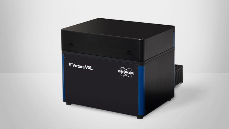
SEE THE LATEST GENERATION VUTARA SMLM SYSTEM:
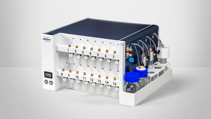
SEE THE LATEST GENERATION MULTIPLEXING ACCESSORY:
References
- Robert Hooke | Biography, Discoveries, & Facts. (2020). Retrieved 1 May 2020, from https://www.britannica.com/biography/Robert-Hooke
- Antonie van Leeuwenhoek | Biography, Discoveries, & Facts. (2020). Retrieved 1 May 2020, from https://www.britannica.com/biography/Antonie-van-Leeuwenhoek
- Robert Koch | German bacteriologist. (2020). Retrieved1 May 2020, from https://www.britannica.com/biography/Robert-Koch
- St. Croix, C., Shand, S., & Watkins, S. (2005). Confocal microscopy: comparisons, applications, and problems. Biotechniques, 39(6S), S2-S5. doi: 10.2144/000112089
- Ebeling, C. (2017). Quantitative Single Molecule Localization Microscopy. Retrieved 27 April 2020, from https://vimeo.com/220672500
- Beyond the diffraction limit. (2009) Nature Photon 3, 361 https://doi.org/10.1038/nphoton.2009.100
- Super-Resolution Imaging of Biological Specimens. (2020). Retrieved 27 April 2020, from https://www.news-medical.net/news/20160927/Super-resolution-imaging-of-biological-specimens-an-interview-with-Dr-Manasa-Gudheti.aspx
- Jungmann, R., Avendaño, M., Woehrstein, J., Dai, M., Shih, W., & Yin, P. (2014). Multiplexed 3D cellular super-resolution imaging with DNA-PAINT and Exchange-PAINT. Nature Methods, 11(3), 313-318. doi:10.1038/nmeth.2835
- DNA-PAINT Imaging with Super-Resolution SML. (2020). Retrieved 20 May 2020, from https://www.bruker.com/products/fluorescence-microscopes/vutara-super-resolution-microscopy/dna-paint-imaging-with-super-resolution-sml.html
- Shivanandan, A., Deschout, H., Scarselli, M., & Radenovic, A. (2014). Challenges in quantitative single molecule localization microscopy. FEBS Letters, 588(19), 3595-3602. doi: 10.1016/j.febslet.2014.06.014
- Szczurek, A., Xing, J., Birk, U., & Cremer, C. (2016). Single Molecule Localization Microscopy of Mammalian Cell Nuclei on the Nanoscale. Frontiers In Genetics, 7. doi: 10.3389/fgene.2016.00114
- Nir, G., Farabella, I., Pérez Estrada, C., Ebeling, C., Beliveau, B., & Sasaki, H. et al. (2018). Walking along chromosomes with super-resolution imaging, contact maps, and integrative modeling. PLOS Genetics, 14(12), e1007872. doi: 10.1371/journal.pgen.1007872
- Vutara Genomics. (2020). Retrieved 27 April 2020, from https://www.bruker.com/products/fluorescence-microscopes/vutara-super-resolution-microscopy/vutara-genomics.html
- Neuroscience | Neurological Disorders | Functional Neuroimaging. (2020). Retrieved 27 April 2020, from https://www.bruker.com/applications/preclinical-imaging/neuroscience.html
- Vutara 352 - Applications | Super-Resolution. (2020). Retrieved 27 April 2020, from https://www.bruker.com/products/fluorescence-microscopes/vutara-super-resolution-microscopy/applications.html
- Milrot, E., Shimoni, E., Dadosh, T., Rechav, K., Unger, T., Van Etten, J., & Minsky, A. (2017). Structural studies demonstrating a bacteriophage-like replication cycle of the eukaryote-infecting Paramecium bursaria chlorella virus-1. PLOS Pathogens, 13(8), e1006562. doi: 10.1371/ journal.ppat.1006562
- Virology Studies with Single Molecule Localization. (2020). Retrieved 20 May 2020, from https://www.bruker.com/products/fluorescence-microscopes/vutara-super-resolution-microscopy/vutara-virology.html
- Alonas, E., Lifland, A., Gudheti, M., Vanover, D., Jung, J., & Zurla, C. et al. (2013). Combining Single RNA Sensitive Probes with Subdiffraction-Limited and Live-Cell Imaging Enables the Characterization of Virus Dynamics in Cells. ACS Nano, 8(1), 302-315. doi: 10.1021/nn405998v
- Tiwari, P., Vanover, D., Lindsay, K., Bawage, S., Kirschman, J., & Bhosle, S. et al. (2018). Engineered mRNA-expressed antibodies prevent respiratory syncytial virus infection. Nature Communications, 9(1). doi: 10.1038/s41467-018-06508-3
- Oncology | Preclinical | MRI | Cancer Treatment. (2020). Retrieved 27 April 2020, from https://www.bruker.com/applications/preclinical-imaging/oncology.html
- Li, Y., Tao, T., Du, L., & Zhu, X. (2020). Three-dimensional genome: developmental technologies and applications in precision medicine. Journal Of Human Genetics, 65(6), 497-511. doi: 10.1038/s10038-020-0737-7
- Hausmann, M., Ilić, N., Pilarczyk, G., Lee, J., Logeswaran, A., & Borroni, A. et al. (2017). Challenges for Super-Resolution Localization Microscopy and Biomolecular Fluorescent Nano-Probing in Cancer Research. International Journal Of Molecular Sciences, 18(10), 2066. doi: 10.3390/ijms18102066
- Nir, G., Farabella, I., Pérez Estrada, C., Ebeling, C., Beliveau, B., & Sasaki, H. et al. (2018). Walking along chromosomes with super-resolution imaging, contact maps, and integrative modeling. PLOS Genetics, 14(12), e1007872. doi: 10.1371/journal.pgen.1007872
- Iuliano, S., Fisher, S. G., Karasik, P. E., Fletcher, R. D., & Singh, S. N. (2002). QRS duration and mortality in patients with congestive heart failure. American Heart Journal, 143(6), 1085–1091. doi: 10.1067/ mhj.2002.122516
- Lichter, J. G., Carruth, E., Mitchell, C., Barth, A. S., Aiba, T., Kass, D. A., … Sachse, F. B. (2014). Remodeling of the sarcomeric cytoskeleton in cardiac ventricular myocytes during heart failure and after cardiac resynchronization therapy. Journal of Molecular and Cellular Cardiology, 72, 186–195. doi: 10.1016/j.yjmcc.2014.03.012
- Live Cell Single Molecule Localization Microscopy. (2020). Retrieved 27 April 2020, from https://www.bruker.com/products/fluorescence-microscopes/vutara-super-resolution-microscopy/live-cell-single-molecule-localization-microscopy.html