Virology Studies
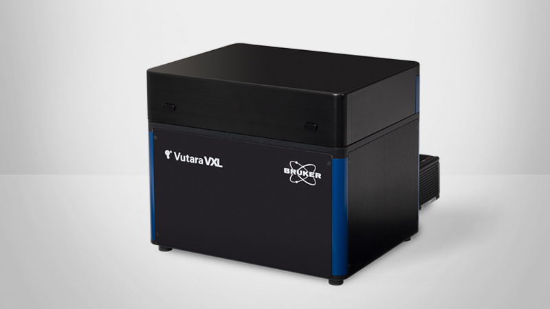
Understanding how viruses behave at different stages of the viral life cycle can reveal key information about disease processes and their real-world implications. The Vutara VXL single-molecule localization microscopy (SMLM) system is uniquely well-suited for studying the mechanisms and cellular consequences of infection, testing possible antiviral therapies, and investigating other questions in virology research.
Vutara VXL is
- The only 3D single-molecule system capable of imaging multiple sample types, from purified virions to tissue sections and whole model organisms;
- Able to deliver high-speed imaging, data acquisition, and analysis for high-quality live imaging and particle tracking studies; and
- Uniquely equipped with integrated multiplexed imaging of the proteome, viral genome, or live-cell samples.
These features have enabled a variety of scientifically critical investigations and are essential for advancing our understanding of viral structure and pathogenesis and virus-host interactions.
To learn more, continue reading, visit the Vutara VXL SMLM microscope page, or contact us directly.
Why Study Viruses with Super-Resolution Microscopy?
Researchers rely on single-particle measurement techniques to analyze the surface composition, interactions, and forces of viral structures. SMLM uniquely allows high-resolution imaging of specific viral components at the molecular level, throughout the entire viral life cycle.
How SMLM creates new avenues for virology research
Conventional techniques like confocal and electron microscopy have struggled to deliver higher resolution images with key information on how different viruses act and move within a cell. No single technique has achieved a resolution sufficient for resolving virus particles while also providing the ability to differentiate molecular components.
Super-resolution microscopy, and specifically single-molecule localization, provides the necessary resolution for imaging the typical size of viruses (20nm to 500nm). Moreover, SMLM delivers a unique combination of multicolor characterization, 3D imaging, and live-cell tracking.
These capabilities make SMLM the best-suited technique for resolving virus particle structural details or determining the localization of virus components within the cellular machinery.
Quantify and Localize Individual Viral Components
SMLM has been shown to be useful for determining viral structure, how viruses attach to target cells, how virus material enters cells, and the effect of viral infection on target cells.
The ability to quantify labeled components in a given target area at the single particle level is a key feature of SMLM. Multicolor localization and 3D particle tracking make it possible to
- Measure the number of proteins and nucleotides in targeted areas of the viral particle and infected cell membrane; and
- Analyze how their distribution changes over time and in response to therapeutic intervention.
The unique ability to perform single-molecule localization of virus samples at both the coverslip and deep within tissue sections has made the Vutara platform the only system capable of imaging virus particle structures, virus particle host cell interactions, and effects of virus infection on cell biology on the same microscope.
As can be seen below, the Vutara VXL is supporting advanced research for:
- COVID-19
- Influenza
- Ebola
- Bovine Alphaherpesvirus 1
- Rhinovirus
Determine Virion Structure
SMLM enables clear visualization and analysis of virion structure and components like surface and internal proteins (spike, membrane, envelope, nucleocapsid) and nucleotides (RNA or DNA).
This technique makes it possible to show details including:
- Virion size and morphology
- The distribution of and spatial relationships between components
The Vutara platform has been used to characterize virion structure, including:
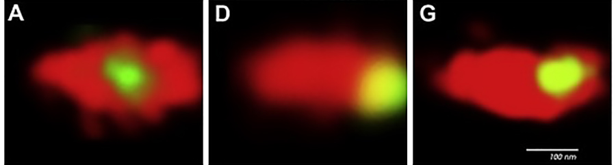
Hodges J., Tang X., Landesman MB., Ruedas JB., Ghimire A., Gudheti MV., Perrault J., Jorgensen EM., Gerton JM., Saffarian S., 2013. Asymmetric Packaging of Polymerases Within Vesicular Stomatitis Virus. Biochemical and Biophysical Research Communications, Volume 440, Issue 2, 271-276. doi.org/10.1016/j.bbrc.2013.09.064.
Using the Vutara SR 200 microscope, Hodges, et al. investigated how different proteins localized throughout the vesicular stomatitis virus (VSV) – an RNA virus with one tapered and one blunt end. SMLM was used to locate the internal proteins within individual virions, proteins were labeled with eGFP, and the envelope of VSV virions was mapped using the same microscopy technique. This enabled the authors to determine the proteins' total fluorescence intensity and the position of their center of fluorescence with respect to the center of the envelope. In doing so, they found that:
- G protein was uniformly distributed on the surface of the virion.
- P and L proteins were packaged asymmetrically at the blunt end of the virion.
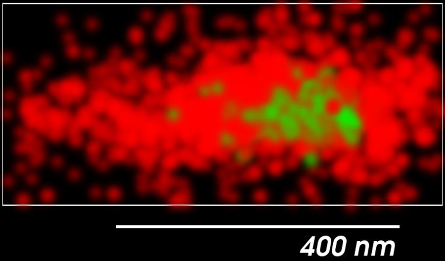
Alonas, E., Lifland, A.W., Gudheti, M., Vanover, D., Jung, J., Zurla, C., Kirschman, J., Fiore, V.F., Douglas, A., Barker, T.H., Yi, H., Wright, E.R., Crowe, J.E., Santangelo, P.J., 2014. Combining Single RNA Sensitive Probes with Subdiffraction-Limited and Live-Cell Imaging Enables the Characterization of Virus Dynamics in Cells. ACS Nano 8, 302–315. doi.org/10.1021/nn405998v
The authors developed MTRIPs (multiply labeled tetravalent RNA imaging probes) -- a method to live label hRSV viral genomes. They then used a Vutara SR 200 and Vutara SRX software to study the early infectivity and replication of enveloped viruses. They were able to identify and isolate individual, infectious, fluorescently labeled viral particles, localize them in three dimensions, and quantify virus replication in single cells over an entire cycle of replication. They found that MTRIP labeling of viral RNA:
- Enabled the simultaneous super-resolution imaging of proteins and the viral genome, which is not possible with conventional fluorescence in situ hybridization techniques (FISH).
- Allows determination of the distribution of viral proteins along the viral gRNA. Only single-molecule localization microscopy gives the resolution to image these sub-300 nm particles.
- Has the potential to become a general methodology for the labeling and study of many important RNA viruses.
Analyze Virus-Host Interactions
The general life cycle of a virus follows the process of attachment, penetration, uncoating, gene expression/replication, assembly, and release.
However, viruses have different methods to undergo viral membrane fusion (entry) and will translocate and express their genome at different rates and manners. Therefore, an in-depth visualization and understanding of how different viruses interact with their host cells, provided by super-resolution microscopy, will give researchers invaluable tools for the effect of these viruses on their hosts.
SMLM allows analysis of a variety of virus-host interactions, such as:
- How viruses move into target cells; and
- The effects of viral infection on target cells, including their intracellular structure, such as cytoskeleton.
The Vutara platform has been used to investigate virus-host interactions, such as:
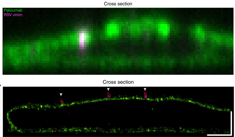
Tiwari, P.M., Vanover, D., Lindsay, K.E., Bawage, S.S., Kirschman, J.L., Bhosle, S., Lifland, A.W., Zurla, C., Santangelo, P.J., 2018. Engineered mRNA-Expressed Antibodies Prevent Respiratory Syncytial Virus Infection. Nature Communications 9, 1–15. doi.org/10.1038/s41467-018-06508-3
The authors used the Vutara microscope to determine the mechanism of action of the therapeutic antibody, palivizumab, on RSV infections.
- Using the Vutara’s ability to image cultured cells in 3D, the authors were able to visualize viral particles on the cell membrane.
- When the cells expressed palivizumab on their cell surface the virions could be observed adjacent, but outside, of the cell membrane (virion size of ~100-300 nm).
- This suggested the mechanism of action of palivizumab is to prevent infection by stopping fusion and cytosolic uptake of RSV.
Milrot, E., Shimoni, E., Dadosh, T., Rechav, K., Unger, T., Etten, J.L.V., Minsky, A., 2017. Structural Studies Demonstrating a Bacteriophage-like Replication Cycle of the Eukaryote-infecting Paramecium Bursaria Chlorella Virus-1. PLOS Pathogens 13, e1006562. doi.org/10.1371/journal.ppat.1006562
The authors used the Vutara to determine the effects of viral infection on cytoskeletal structure. From this, they were able to determine that the actin cytoskeleton plays a critical role in viral infectivity.
- The authors used super-resolution imaging to monitor how the microtubule and actin cytoskeleton changed over the course of viral infection.
- During infection, the microtubule network became more fragmented and disappeared from the center of the cell.
- During infection, the actin cytoskeleton loses its fine structure at the periphery of the cell and forms a shell around the rounded edge of the cell.
- Pharmacological experiments and other experiments revealed that disruption of the microtubule network had little effect on virion production, while disruption of the actin cytoskeleton reduced virion production.
Akiyama H., Ramirez NGP., Gudheti MV., Gummuluru S., 2015. CD169-Mediated Trafficking of HIV to Plasma Membrane Invaginations in Dendritic Cells Attenuates Efficacy of Anti-gp120 Broadly Neutralizing Antibodies. PLOS Pathogens 11(3): e1004751. doi.org/10.1371/journal.ppat.1004751
Myeloid dendritic cells (DCs) are important to elicit a potent antiviral immunity and can capture HIV-1 via the receptor CD169/Siglec-1 that binds to the ganglioside, GM3, in the virus particle membrane. This research group utilized the Vutara 200 microscope, to investigate the role of CD169 in HIV-1 trafficking and trans-infection. Using super-resolution microscopy, they revealed:
- In a LPS induced cell, CD169 and HIV-1 accumulated in pocket-like compartments.
- LPS and IFN-α can both upregulate CD169 expression.
- HIV virions were closely associated with CD169.
Monitor Infection Dynamics
Bruker’s super-resolution fluorescence microscopy solutions are unique in their ability to provide the tools, resolution, and post-processing ability to capture the progression and distribution of infection for a variety of experiments.
The Vutara system has been used to analyze:
- Early stages of infection.
- Spatio-temporal investigation of viral replication/propagation of infection.
- Lytic or persistent infection (quantify the abundance of free viruses released during cell lysis).
- Distribution of infected cells in their tissue environment.
The Vutara platform has been used to investigate a variety of infection dynamics, such as:
Alonas, E., Lifland, A.W., Gudheti, M., Vanover, D., Jung, J., Zurla, C., Kirschman, J., Fiore, V.F., Douglas, A., Barker, T.H., Yi, H., Wright, E.R., Crowe, J.E., Santangelo, P.J., 2014. Combining Single RNA Sensitive Probes with Subdiffraction-Limited and Live-Cell Imaging Enables the Characterization of Virus Dynamics in Cells. ACS Nano 8, 302–315. doi.org/10.1021/nn405998v
The authors developed MTRIPs (multiply labeled tetravalent RNA imaging probes) -- a method to live label hRSV viral genomes. They then used a Vutara SR 200 and Vutara SRX software to study the early infectivity and replication of enveloped viruses. They were able to identify and isolate individual, infectious, fluorescently labeled viral particles, localize them in three dimensions, and quantify virus replication in single cells over an entire cycle of replication. They found that MTRIP labeling of viral RNA:
- Enabled the simultaneous super-resolution imaging of proteins and the viral genome, which is not possible with conventional fluorescence in situ hybridization techniques (FISH).
- Allows determination of the distribution of viral proteins along the viral gRNA. Only single-molecule localization microscopy gives the resolution to image these sub-300 nm particles.
- Has the potential to become a general methodology for the labeling and study of many important RNA viruses.
Yaakov, L.B., Mutsafi, Y., Porat, Z., Dadosh, T. and Minsky, A., 2019. Kinetics of Mimivirus Infection Stages Quantified Using Image Flow Cytometry. Cytometry, 95: 534-548. doi.org/10.1002/cyto.a.23770
In this study, the authors investigated the temporal progression of events in virally infected cells using the Vutara 200 microscope. Motivated by the ability to analyze large populations of infected cells despite the heterogeneity of viruses and their hosts, they utilized flow cytometry to study Mimivirus and Acanthamoeba polyphaga. Their work found:
- The microtubule network became more fragmented and disappeared from the center of the cell.
- The actin cytoskeleton lost its fine structure at the periphery of the cell and formed a shell around the cell edge.
Image Live Cells
Live-cell imaging is a powerful tool for visualizing the dynamics of viral particles in living cells
Virus researchers may also be interested in the live cell and single-molecule particle tracking capabilities of the Vutara platform. The Vutara VXL is fully capable of both live-cell single-molecule imaging of cellular structures, such as organelles, and single-molecule particle tracking. Uniquely, the Vutara is also capable of combining these two techniques to do particle tracking in conjunction with cellular structure imaging.
Please refer to the live cell imaging webpage and the Vutara live cell imaging webinar to learn more about live cell imaging with the Vutara microscope and SRX software.
Develop Safer and More Effective Therapeutic Interventions
SMLM can provide a holistic picture of the behavior of viruses, their mechanisms of infection and transmission, and the cellular consequences of infection. This information enables researchers to develop better intervening measures against viruses, which can be used to prevent and control the spread of disease. SMLM has also been instrumental in testing therapeutic interventions for both efficacy and safety.
Specific target cell membrane proteins involved in infection can be identified, and functional significance as well as possible therapeutic strategies can be explored.
The Vutara platform has been used to investigate prophylactic antivirals, including their:
Tiwari, P.M., Vanover, D., Lindsay, K.E. et al. Engineered mRNA-expressed antibodies prevent respiratory syncytial virus infection. Nat Commun 9, 3999 (2018). doi.org/10.1038/s41467-018-06508-3
This research group used the Vutara 352 microscope and Vutara SRX software to evaluate a novel mRNA-based approach to preventing respiratory syncytial virus (hRSV) infection -- and potentially other viral infections -- in the lung. Using the Vutara for both widefield and super-resolution imaging, the authors were able to (1) investigate the expressed antibodies' methods for preventing infection and (2) test the intervention for cellular consequences, like undesirable inflammation, that could present safety concerns for its translational application. They found:
- mRNA-expressed anchored neutralizing antibodies are retained on the plasma membrane of transfected cells.
- mRNA-based expression of neutralizing antibodies can prevent RSV infection in vivo and in vitro.
- Delivery of mRNA-expressed antibodies caused no significant inflammatory response.
- Data suggests that expressing membrane-anchored broadly neutralizing antibodies in the lungs could be a promising pulmonary prophylaxis approach.
- Theoretically, many expressed therapeutic proteins could be anchored and delivered in vitro in this manner.
Still have questions about SMLM for virology research? Contact our super-resolution microscopy experts to discuss your unique research needs.
Vutara Single-Molecule Localization Microscope for Viral Research
The Vutara single-molecule localization system has been a critical instrument for understanding viral particles.
Specifically, the Bruker Vutara VXL provides:
- The resolution needed for the typical size of viruses (20nm to 500nm).
- Propriety biplane technology that achieves at least 20 nm XY and 50 nm Z precision.
- High-speed acquisition -- ideal for live imaging, particle tracking, and rapid data acquisition.
- Integrated fluidics for multiplexed imaging of the proteome, genome, or live-cell applications.
- Powerful acquisition software with real-time single-molecule localization.
- Powerful visualization and analysis software package provides a complete statistics tool set.


Frequently Asked Questions
SMLM microscopy allows labeling and imaging of viral components such as proteins (spike, membrane, envelope, nucleocapsid) and nucleotides (RNA or DNA) at a resolution that allows clear visualization and analysis. EM can achieve a resolution sufficient to resolve viruses, but differentiation of molecule components is not possible. Diffraction-limited techniques such as confocal microscopy allow fluorescent labeling of components but cannot resolve sub-diffraction components. SMLM can.
SMLM allows analysis of a variety of virus-host interactions. SMLM can allow visualization and analysis of specific target cell membrane proteins involved in viral infection. Specific target cell membrane proteins involved in infection can be identified, and functional significance as well as possible therapeutic strategies can be explored. Conversely, SMLM allows investigation of the role of specific viral proteins in the infection process. SMLM allows analysis of how viruses move into target cells, primarily through single particle tracking. SMLM also allows analysis of the effect of viral infection of target cells by imaging at the super-resolution level changes in target cell intracellular structure, such as cytoskeleton.
Challenges to imaging viruses with SMLM are primarily related to developing labeling protocols for imaging viral and target cell components.
SMLM allows imaging and analysis at the molecular level of viral components and target cell components involved in viral infection. As with most biological research, SMLM will not be the only technique used to study viruses and the impact of viral infection on target cells. Other techniques, such as sequencing to determine nucleotide sequences and viruses, as well as assays to evaluate target cells such as proteomics and assays of metabolism, will continue to be employed.
SMLM allows researchers to label and image molecular components of viruses and target cells at the super-resolution level. SMLM has been shown to be useful to determine viral structure, how viruses attach to target cells, how virus material enters cells, and the effect of viral infection on target cells.
SMLM provides a tool for better understanding of the mechanisms of viral infection as well as a tool ideally suited to explore therapy strategies at the molecular level.