SCiLS™ Software Solutions
Turn data into knowledge
SCiLS™ software - comprehensive and easy to use


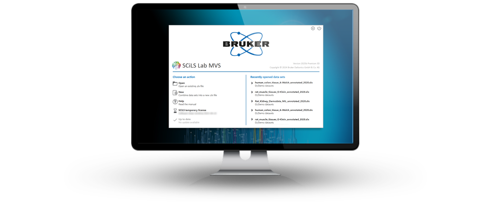
SCiLS™ Software
Software for MALDI Imaging data analysis
Analysis and visualization of MALDI Imaging data has never been easier.
Powerful visualization and reporting
- Explore every feature in every spectrum
- Study molecular images directly in histopathological context
- Compelling statistical charts and reports
- Volumetric visualization of 3D MS imaging studies (optional)
Advanced data processing and analysis
- Advanced supervised and unsupervised machine learning algorithms
- Comparative analysis to uncover discriminative and correlated features
- Spatial segmentation and component analysis for untargeted clustering
- Metadata annotations for efficient region characterization and grouping
- Classification models for categorization of unlabeled samples
- Analyte quantitation based on dilution series
Workflow and extensibility
- Comprehensive tools for targeted and untargeted SpatialOMx®
- Integration with histological annotations from digital pathology
- Extensive application programming interface (API) for automated reporting and advanced workflow integration
- Export MS imaging data to the open industry standard imzML
- Import MS imaging datasets from imzML or from various third party vendors’ imaging data formats (optional)
- Integrated molecular annotation powered by MetaboScape® (optional)
- Integrated iprm-PASEF workflow to identify ion images with confidence
- Explore spatial multiomics data using the SCiLS™ Ion Image Mapper
System requirements
The following system requirements are recommended:
- Intel® Core™ i7 processor or Intel® Xeon® processor (or equivalent) with at least 4 cores
- Microsoft® Windows® 10 or 11 (64-Bit versions only, minimum Windows® 10 version 1607 or later)
- 64 GB of RAM (128 GB recommended for large datasets)
- 1 TB disk space (SSD recommended, see below)
- Full-HD display (1920 x 1080) or larger
- Graphics adapter supporting OpenGL 3.2, at least 4 GB GPU memory recommended
For best performance, a solid state drive (SSD) should be used to host the SCiLS™ Lab data files.
A new and easy-to-use platform for sharing and viewing
SCiLS™ Scope lowers the barriers for sharing and viewing MALDI Imaging and MALDI HiPLEX-IHC data. With an easy-to-use, lightweight interface and data in the open OME-TIFF format, it is ideal for sharing data between collaborators who want to focus on examining ion images. Review and analyse ion images intuitively by adjusting their brightness and contrast, overlaying individual channels, and measure distances between biologically relevant structures.
The software fits seamlessly into the SCiLS™ autopilot workflow, which automates data acquisition, importing data into SCiLS™ Lab and exporting predefined target ion images into the OME-TIFF format. A ready-to-go solution for MALDI Imaging and MALDI HiPLEX-IHC.
World-leading software for analysis of MALDI Imaging data
SCiLS™ Lab integrates data from all major mass spectrometry vendors, including Bruker’s FLEX series and MRMS, as well as datasets provided in the open imzML format (optional). For timsTOF fleX ion mobility data, SCiLS™ Lab displays mass-mobility ion images allowing CCS-enabled data interpretation.
Comparative analysis of multiple samples can be visualized in both 2D and 3D, enabling a multitude of applications in pharmaceutical drug development, tissue-resolved biomarker discovery, and translational pathology research to complement immunohistochemistry.
SCiLS™ Lab supports intuitive workflows for MALDI Imaging quantitation of target molecules directly from tissue. It can interpret MALDI MS/MS Imaging data generated using iprm-PASEF® and integrates with MetaboScape® for the molecular annotation of compounds with confidence. The SCiLS™ Ion Image Mapper allows easy fusion of MALDI Imaging-based spatial multiomics data. For advanced histopathological analysis, SCiLS Lab provides an interface to the powerful open source software QuPath, widely used in digital pathology.
Label-free MALDI Imaging for all classes of molecules – from proteins to small molecules
SCiLS™ Lab can be used for the processing and analysis of multiple MALDI Imaging datasets, allowing them to be loaded and arranged side by side. Compositions of data sets of virtually unlimited size can be visualized and statistically analyzed for detailed interpretation. SCiLS™ Lab can be used to generate and visualize 3D models from consecutive 2D MALDI Imaging datasets. Moreover, the SCiLS™ Ion Image Mapper can be used to fuse spatial multiomics data sets which can be jointly analyzed in SCiLS™ Lab, providing unparalleled actionable knowledge.
Large sample cohorts in clinical research studies
In clinical biomarker studies for the discovery of discriminating ions, large sample cohorts and huge amounts of data need to be analyzed. SCiLS™ Lab makes it easy to link sample metainformation to MALDI MSI data, saving time and avoiding errors. Histological annotations from digital pathology are integrated using an interface to the powerful and widely used open source application QuPath.
Automatic elucidation of spectral features discriminating pathophysiological alterations are easily performed. With just one click, SCiLS™ Lab automatically quantifies how well ions discriminate between different biological states. The resulting list of novel biomarker candidates can be easily exported for subsequent analysis.
Discover the powerful data mining capabilities that are now just a single click away!
Whole body imaging
The visualization of drug and metabolite distributions within whole-body tissue sections remains a challenging methodology with respect to sample processing, acquisition, and computational data analysis.
SCiLS™ Lab offers tools for the entire MALDI MSI data analysis workflow. Multiple data sets composed of millions of spectra can be processed using one software, from the registration of multiple individual data sets over spectral preprocessing to comprehensive statistical analysis.
"SCiLS Lab transforms MALDI Imaging workflows — whether for N-linked glycans, extracellular matrix peptides, or both — combining ease of use, multiplex scalability, and compatibility with timsTOF fleX and MRMS, to drive discovery across large clinical and translational study cohorts."
Peggi Angel, Ph.D., Professor, Department of Pharmacology & Immunology, Medical University of South Carolina
"The multiomics and multimodal data fusion and analysis capabilities of SCiLS Lab were a great help integrating and analyzing a record-breaking eight sequential MALDI Imaging acquisitions from a single tissue."
Erin Seeley, Ph.D., Director, Mass Spectrometry Imaging Core, MD Anderson Cancer Center
"SCiLS Lab has been an essential tool in our research. Its integration with QuPath allows us to seamlessly combine the histomorphological insights from our pathologists with the detailed, feature-rich, spatially resolved molecular data from MALDI Imaging, enhancing both interpretation and discovery."
Juliana Gonçalves, Ph.D., Research Assistant, Insitute of Pathology, School of Medicine and Health, Technical University of Munich, Munich, Germany
"With SCiLS™ Lab our team saved endless hours of manual comparisons. This unique feature was eagerly anticipated by us."
Rita Casadonte, Ph.D., Research Scientist/Group Leader at Proteopath GmbH, Trier, Germany
For Research Use Only. Not for use in clinical diagnostic procedures.