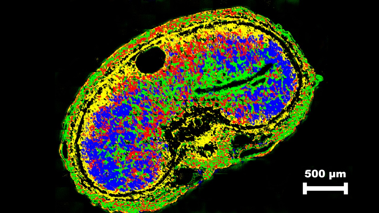
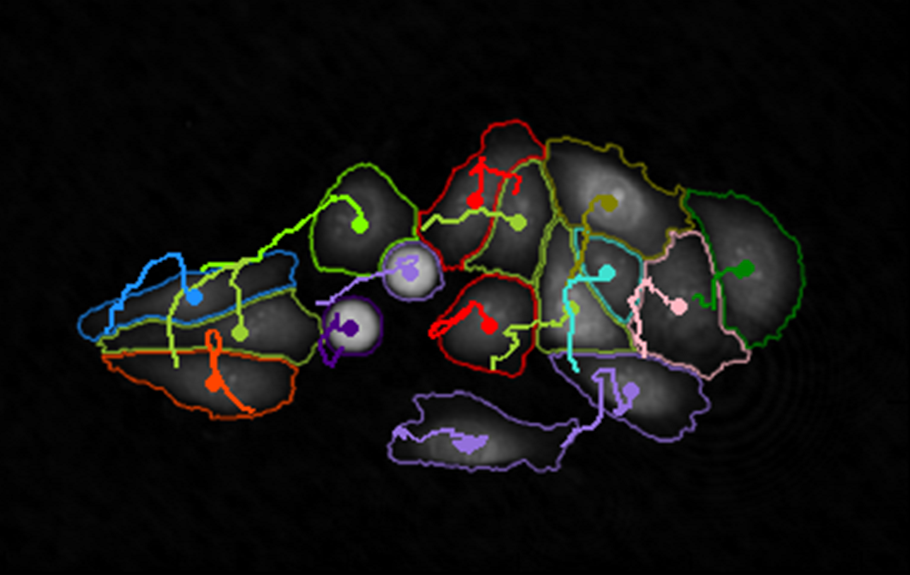
It is when biological tissues lack contrast in X-ray absorption allowing visualization by microCT. Conventional X-ray imaging (leaving aside for now phase contrast techniques) requires different structural components to have differing X-ray absorption for structures to be contrasted; microCT is no exception. Figure 1 shows the difference between microCT images of a mouse embryo obtained without staining (a) and after staining in phosphotungstic acid or “PTA” (b).
Whereas in microscope histology stains confer visual color, for microCT stains – or “contrast agents” – confer X-ray contrast. Thus the stain must contain a heavy element with high atomic number (Z) for strong X-ray attenuation. Tungsten in PTA (Z=74) fits the bill for this. Stains must also have differential tissue specificity. Some classic histology stains also work for microCT. PTA is an example; it has long been used as both fixative and histology stain [i, ii, iii] for collagen and fibrin. Metscher [iv] has showed PTA also to be an effective microCT stain. His paper has become a classic reference for microCT study of soft tissue with its helpful instructions on use of PTA and other contrast agents (such as iodine). PTA can be used dissolved either in water or in ethanol. Lendrum et al. [v] observed “that a water-soluble dye (e.g. PTA) dissolved in alcohol then behaves as if it were of a smaller molecular size.” Thus when in ethanol PTA may pervade into finer tissue structures. The group of Dr Mary Barbe in Temple University, Philadelphia, USA have added important refinements, such as to thoroughly wash out buffer (with water) from samples prior to staining to ensure optimal tissue pervasion of PTA [vi, vii].
Staining mouse embryos with PTA can be found in the method note “MN007_Embryo staining with PTA for exvivo micro-ct imaging”. Beyond PTA, Lugol’s iodine is also an effective microCT stain; Degenhardt [viii] carefully optimized this method to maximize penetration while minimizing shrinkage. They found that “staining with 25% Lugol for 48 hours resulted in complete and uniform tissue penetration with minimal shrinkage”