Light-Sheet Images of Organoids and Cell Cultures
Organoids and 3D Cell Cultures
Cell cultures are an important tool in life sciences to study biological processes in a controlled set up. In recent years, growing cells in a 3D environment, rather than 2D, has become increasingly important. These environments typically use a gel-like matrix or scaffold that mimics the extracellular matrix found in tissues.
Approaches used in laboratory settings are 3D cell cultures, organoids, and spheroids. Their origins range from primary cell cultures from different organisms to tumors or stem cells. These cells are useful tools as they are genetically modified with intrinsic tags and targets.
- 3D cell culture is a generic term referring to the cultivation of cells in a 3D environment that closely resembles natural tissues or organ architecture.
- Spheroids are 3D cell aggregates that form when cells self-assemble into spherical structures. Spheroids can be generated through various techniques, lack a defined tissue architecture, and often have a random cell distribution.
- Organoids are 3D cell cultures that closely mimic the characteristics of specific organs or tissues. They exhibit a high level of cellular organization, are derived from stem cells or primary tissue samples, and are cultured in conditions that promote self-organization. Organoids are more complex than spheroids and exhibit tissue-specific organization, including multiple cell types and physiological functions.
3D cell culture is a general term used when cells are grown in a three-dimensional environment, while spheroids are simpler 3D cell aggregates, and organoids are more complex. The choice of approach depends on the research objectives and the complexity level needed for experiments.
Types of 3D Cell Culture Imaging
While live time-lapse imaging remains a prominent application in 3D cell culture imaging, the significance of tissue perturbation applications is steadily increasing. This includes applications such as laser ablation for biophysical studies and uncaging or caging photomanipulation. In addition to live imaging, 3D cell cultures can also be fixed, stained, or prepared with tissue clearing, expansion, or sectioning for subsequent imaging.
3D cell cultures enable:
- Understanding Development: Researchers study the development of organs and tissues in previously impossible ways, shedding light on developmental biology and congenital diseases.
- Personalized Medicine: Organoids can be stem-cell derived organoids from patient cells, allowing the establishment of personalized treatments tailored to the patient.
- Cancer Research: Organoids can be used to model different cancer types, facilitating the discovery of new cancer therapies and the study of drug resistance mechanisms.
Why light-sheet imaging for 3D cell cultures?
3D cell cultures are fragile systems that require optimized experimental protocols and low phototoxicity during imaging to preserve samples. Light-sheet imaging is a unique solution for these challenges by enabling researchers to study 3D cell culture systems, including time-lapse imaging.
Bruker's light-sheet systems were designed with scientists to ensure sample health, ease of use, and high imaging quality. Every system is suitable for 3D cell culture work.
3D Imaging of Organoids
Unlike traditional 2D cell cultures, organoids are complex 3D tissue models with tissue-like structures that require advanced imaging methods to capture their intricate architecture.
One widely used approach for 3D imaging of organoids is light-sheet microscopy. This enables researchers to construct detailed 3D images of organoids, revealing the spatial organization of cells, tissue layers, and organoids.
Advanced imaging methods not only enhance our understanding of organoid biology, but also contribute to the optimization of organoid culture protocols. Furthermore, insights into the behavior and morphology of organoid models in a native 3D context contribute to the development of more effective therapies.
Applications include:
- Disease modeling
- Drug screening
- Personalized medicine
- Regenerative medicine
- High-throughput screening

3D Cell Culture
3D culture system of human primary cells imaged on the QuVi SPIM (330µm x 220 µm x 1200µm).
Courtesy of:
Yassen Abbas, Turco Lab, University of Cambridge, Cambridge, UK

HeLa Cell Culture
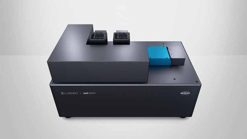
HeLa cells expressing GFP and mCherry. Imaged on the InVi SPIM.
Courtesy of:
Tobias A. Knoch, Erasmus MC, Rotterdam, The Netherlands.

Pancreatic Sphere
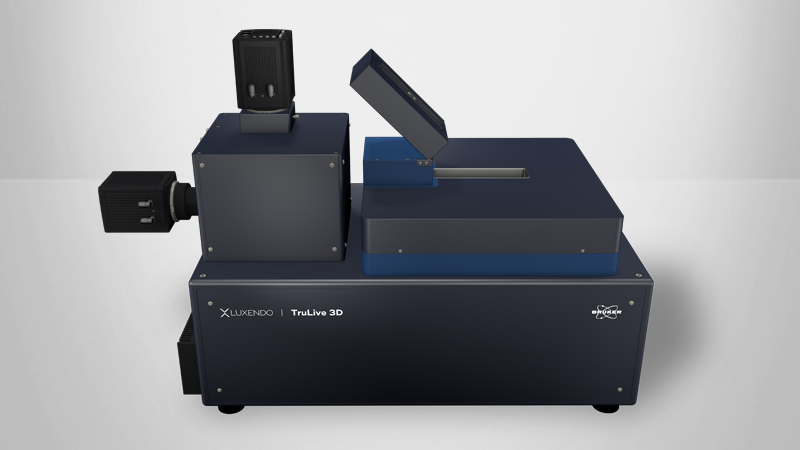
hESC-derived pancreatic spheres. Imaging on the TrueLive3D Imager enables collecting information of several samples in one experiment. Visualization: Imaris (Bitplane).
Courtesy of:
Yung Hae Kim, Graphin-Botton Group, MPI-CBG, Dresden, Germany
Pancreatic Sphere
hESC-derived pancreatic spheres. Imaging on the TruLive3D Imager enables collecting information of several samples in one experiment. Visualization: Imaris (Bitplane)
Courtesy of:
Yung Hae Kim, Graphin-Botton Group, MPI-CBG, Dresden, Germany
Tumorigenesis in Mammary Organoids
Characterization and imaging of stochastic tumorigenesis in mammary organoids. Imaged on the InVi SPIM.
A. Alladin, L. Chaible, L. Garcia del Valle, S. Reither Sabine, M. Loeschinger, M. Wachsmuth, J.K. Hériché, C. Tischer, M, Jechlinger. Tracking cells in epithelial acini by light-sheet microscopy reveals proximity effects in breast cancer initiation. eLife 2020;9:e54066 doi: 10.7554/eLife.54066
Fixed Astrocyte Spheroid
Spheroid stained with anti-GFAP (Alexa 488) to label astrocytes and anti-Neurofilament200 (Alexa555) to label neurons. Imaged on the InVi SPIM.
Courtesy of:
Markus Bruell, AG Leist, University of Konstanz, Konstanz, Germany
Case Study
In a study by Zilova, L., Weinhardt, V., et al (2021), retinal tissue differentiation and development are examined in fish primary embryonic pluripotent cells. Using the Acquifer IM, the group studied the processes of aggregation and compaction in cellular aggregates.

In this same study, Bruker's Luxendo MuVi light-sheet microscope was used to investigate the 3D structure and interior composition of organoids.


Live Imaging of Cell Cultures
Live-cell imaging focuses on observing living cells over time. This technique involves the use of environmental controls to maintain optimal conditions for cell viability during imaging, including temperature, humidity, and CO2 levels.
Time-lapse imaging is invaluable for studying dynamic cellular processes such as cell motility, intracellular trafficking, and cell signaling. It also plays a critical role in drug discovery and development, as it allows for the real-time assessment of drug effects on cell behavior.
The combination of cell culture and advanced imaging technologies continues to drive groundbreaking discoveries in fields such as cancer research, neuroscience, and regenerative medicine by providing insights into the inner workings of cells and their responses to various stimuli.
Cell Culture Imaging
Mitosis in HeLa Cells
Mitosis in HeLa cells stained for histone 2B-mCherry (magenta), GFP-tubulin (green) and GFP-tubulin (white, deconvolved).
Imaged on the InVi SPIM Lattice Pro.
Visualization: Imaris (Bitplane).
Courtesy of:
Sabine Reither
European Molecualr Biology Laboratory (EMBL)
Heidelberg, Germany
Sample: HeLa cells (Neumann et al., Nature. 2010 Apr 1;464(7289):721-7)
Mouse Pre-implantation Development
Left: Mouse preimplantation embryos expressing H2B-mCherry. Nuclei tracking from one-cell stage to blastocyst.
Right: Mouse oocytes expressing CENPC-EGFP and H2B-mCherry for kinetochore tracking.
Imaged on the InVi SPIM.
Petr Strnad, et al. (2016). Inverted light-sheet microscope for imaging mouse pre-implantation development. Nature Methods 13, 139-145
3D Cell Culture
3D culture system of human primary cells imaged on the QuVi SPIM.
Courtesy of:
Yassen Abbas
Turco Lab, University of Cambridge
Cambridge, UK

Epithelial Cells
Epithelial cell line (BS-C-1) stained for f-actin and chromatin. Image taken with InVi SPIM Lattice Pro.
Courtesy of:
Ulrike Engel
Nikon Imaging Center
University of Heidelberg
Germany

HeLa Cell Culture
HeLa cells expressing GFP and mCherry. Imaged on the InVi SPIM.
Courtesy of:
Tobias A. Knoch
Erasmus MC
Rotterdam, The Netherlands
Light-Sheet Publications with Organoids
| Year | Journal | Title | Author(s) | Imaging Platform | Subject | Methodology |
|---|---|---|---|---|---|---|
| 2024 | Distinct tumor architectures and microenvironments for the initiation of breast cancer metastasis in the brain | Gan et al | MuVi SPIM | TNC spheroid, microglia, tumour response, perivascular, brain metastasis | cell lines, mouse, immunohistochemistry, Oncosphere culture, In vitro cell proliferation assay, In vitro wound scratch assay, CRISPRi | |
| 2022 | Nature | Periodic formation of epithelial somites from human pluripotent stem cells | M Sanaki-Matsumiya, M Matsuda, N Gritti… Miki Ebisuya | MuVi SPIM | embryonic development, somitogenesis, segmentation clock | Human iPSC cultures, human somitoids, human somitoids, HCR, IHC, timelapses, human somitoids, morphometry |
| 2022 | The Lancet | Müller glia fused with adult stem cells undergo neural differentiation in human retinal models | SÀ Bonilla-Pons, S Nakagawa, EG Bahima… Maria Pia Cosma | MuVi SPIM | retina organoids, Müller glia, regeneration | organotypic cultures of human retina, preparations of dissociated cells, microinjection system, qRT-PCR, immunofluorescence, Flow cytometry |
| 2022 | STEM CELLS AND REGENERATION | Rapid and robust directed differentiation of mouse epiblast stem cells into definitive endoderm and forebrain organoids | D Medina-Cano, EK Corrigan, RA Glenn… Thomas Vierbuchen | TrueLive3D Imager | developmental biology, organoids | mouse, pluripotent stem cells, Wnt pathway inhibitors, definitive endoderm and neural organoids,differentiation models |
| 2021 | eLife | Fish primary embryonic pluripotent cells assemble into retinal tissue mirroring in vivo early eye development | L Zilova, V Weinhardt, T Tavhelidse, C Schlagheck… Joachim Wittbrodt | MuVi SPIM | morphogenesis and differentiation | medaka and zebrafish, embryonic pluripotent stem cells, fluorescent labeling, quantification |
| 2021 | Cell Reports | Endothelial Wnts control mammary epithelial patterning via fibroblast signaling | J Wang, W Song, R Yang, C Li, T Wu, XB Dong, B Zhou… Yi Arial Zeng | MuVi SPIM | endothelial cells, Wnt, fibroblasts | fluorescent mouse, organoid, FACS, immnohistochemistry, RNA-seq |
| 2020 | Elife | Tracking cells in epithelial acini by light sheet microscopy reveals proximity effects in breast cancer initiation | A Alladin, L Chaible, L Garcia del Valle, R Sabine… Martin Jechlinger | InVi SPIM | cancer environment, oncogenes, breast cancer | mouse, 3D acinus cultures, qPCR analysis, image analysis, |
| 2020 | ALTEX | Incorporation of stem cell-derived astrocytes into neuronal organoids to allow neuro-glial interactions in toxicological studies | M Brüll, AS Spreng, S Gutbier, D Loser, A Krebs… | MuVi SPIM | neuro-glial interactions , astrocytes, organoids | cell culture, proliferation, imaging |