Languages

nanoIR Spectroscopy Journal Club

Infrared Nanospectroscopy Reveals the Molecular Interaction Fingerprint of an Aggregation Inhibitor With Single Aβ42 Oligomers

by F. S. Ruggeri, J. Habchi, S. Chia, R. I. Horne, M. Vendruscolo, and T. P. J. Knowles

Key Points

  • The single molecule morphological and chemical sensitivity of infrared nanospectroscopy facilitates the first direct measurement of the structure and interaction between single Aβ42 protein and an aggregation inhibitor, bexarotene; and
  • The formation of a single hydrogen bond between bexarotene and Aβ42 inhibits Aβ42 aggregation, which reveals important information about the molecular mechanisms responsible for protein aggregation.


 Receive future Journal Club updates via email.

This review appeared in the February 2022 edition of the nanoIR Journal Club — a monthly email brief highlighting leading-edge research and the latest discoveries supported by Bruker nanoIR technology.

Nature Communications 12, 688 (2021)
DOI: 10.1038/s41467-020-20782-0

The misfolding and subsequent aggregation of amyloid-β (Aβ) proteins is believed to play a significant role in the development of medical conditions, such as Alzheimer’s disease. Despite significant efforts to develop compounds that can interfere with aggregation pathways of proteins related to misfolding disorders, no such disease-modifying drug has been developed to date. One of the main reasons for this is our incomplete knowledge of the molecular mechanisms underlying the process by which small molecules interact with proteins and interfere with their aggregation. Thus, it is very desirable to employ new analytical approaches to help us understand the molecular mechanism responsible for such processes.

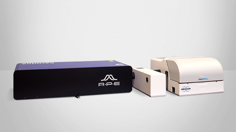
In this work, the authors used Bruker's nanoIR2 nanoscale IR spectroscopy system to perform the first direct measurement of the molecular interaction between single Aβ42 oligomeric and fibrillar species and a small molecule aggregation inhibitor, bexarotene. Their work elucidates how this small molecule interacts with the proteins and interferes with their aggregation pathways. This approach shows great promise for providing insights leading to the discovery of structure-based drugs for protein misfolding diseases.


Learn more about this study

In this study, the aggregation reaction of Aβ42 was investigated in the absence and the presence of bexarotene and one of its derivatives, where the carboxyl group was substituted by an ester functional group. AFM-IR spectra were collected from the Aβ42 aggregates at the single protein molecule scale. Second derivative analysis was then applied to deconvolve the amide I band of the aggregates to evaluate their secondary structures. Compared to the oligomeric aggregates, the fibrillar aggregates showed increased absorption at 1630 cm⁻¹, indicating a conversion from antiparallel to parallel β-sheet conformation in the fibrilla. In the presence of bexarotene, AFM-IR spectrum of the aggregates indicated that the peak wavenumber of C=O stretching band of bexarotene occurred at 1725 cm⁻¹, a significant blue shift relative to its location in pure bexarotene at 1690 cm⁻¹. This chemical shift demonstrates that bexarotene is transitioning from a dimeric form with double coordination to a monomeric form coordinating with the amyloid aggregates through hydrogen bonding. The derivative of bexarotene that contains a methyl ester group did not affect the Aβ42 aggregation, as its methyl ester group did not form a strong hydrogen bond with the Aβ42 aggregate.

Overall, results from this work showed that the small molecule of bexarotene is able to inhibit the Aβ42 aggregation through the formation of a single hydrogen bond between bexarotene and Aβ42. This work also demonstrated that AFM-IR is a powerful technique for helping to elucidate molecular mechanisms in biological processes, such as protein folding.


      KEY TERMS:

  • AFM-IR; Infrared Nanospectroscopy; Drug Discovery; Molecular Conformation; Protein Aggregation, Single-Molecule Biophysics