Fundamental Research
Brain phenotyping advances fundamental neurological understanding
IResearchers in the field of fundamental neuroscience study the nervous system to understand its structure, function and molecular biology during development under both normal and pathological conditions.
Neuroscientists apply medical imaging methods such as magnetic resonance imaging (MRI), functional MRI (fMRI), positron emission tomography (PET) and magnetic resonance spectroscopy (MRS) in preclinical studies to learn about the brain’s impact on behavior and cognition. This helps them to understand what happens to the nervous system in healthy processes, as well as in cases of neurological disorders such as Alzheimer’s, Parkinson’s, stroke, memory loss and cognitive decline.
Some of the basic research is potentially translatable such as studies into stem cell therapies that aid nerve regeneration or the development of materials that can promote repair in spinal cord injuries. Scientists in both academia and industry agree that fundamental research is crucial for advancing the clinical area.
Bruker BioSpec users investigate anatomical structures, pathologies, and metabolic anomalies in the rodent brain. The BioSpecs also offer fMRI for the analysis of brain function, enabling researchers to detect active brain areas and visualize what happens when the brain is thinking. To meet researchers need to identify smallest details, Bruker’s ultra-high field BioSpec MRI instruments can be even further enhanced with state-of-the-art MRI CryoProbe technology.
For brain metabolism studies, researchers can use Bruker’s NMR spectroscopy instruments to visualize metabolic exchanges that take place between astrocytes and neurons, for example. Furthermore, MR/PET hybrid modalities synchronize the detailed, soft-tissue imaging that MRI affords with the metabolic imaging capabilities of PET.
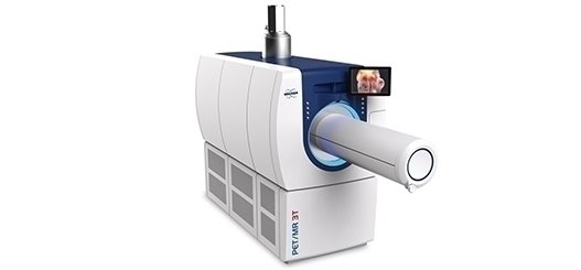
To perform molecular phenotyping of brain tissue and characterize different glial cells, for example, neuroscientists can use Bruker’s Ultraflex II MALDI-TOF/ TOF mass spectrometer. Furthermore, the BioSpec 3T provides an ideal solution for morphological animal phenotyping.
Researchers are using the simultaneous holographic photostimulation and imaging in vivo capabilities of our Ultima multiphoton microscopes to understand links between neural circuit patterns of activity and behavior in awake-behaving animals.