Fluorescence Microscopy Journal Club

Single-Molecule Tracking of Chromatin-Associated Proteins in the C. elegans Gonad

by Lexy von Diezmann and Ofer Rog

Key Points

  • PAJF549 offered greater photostability and higher photon output than the photoconvertible protein mMaple3, resulting in longer, more robust tracking;
  • Diffusive proteins appear to avoid the nucleolus, selectively exploring chromatin‑rich regions, and
  • When combined with well-established gonad extrusion protocols, improved single‑molecule tracking strategies are well-suited for investigating chromosome organization and studying the molecular dynamics associated with this process.


 Receive future Journal Club updates via email

The Journal of Physical Chemistry B, 2021 125 (23), 6162-6170
DOI: 10.1021/acs.jpcb.1c03040

The nucleus is responsible for containing the genomic information of a cell and is highly organized in order to perform a range of functions relating to cell division, transcription, and ribosome biogenesis. Of particular interest to developmental biologists is meiosis, the cell‑division cycle responsible for the production of gametes. During this process, parental chromosomes break and repair in a unique and highly organized fashion through a series of loops on a meiotic axis. This organization is essential to the proper partitioning of chromosomes into gametes. Caenorhabditis elegans is a biological system that is well-suited for studying this process and observing protein interaction with intact chromosomes within their native environment due to their temporally ordered gametes and well-separated chromosomes within meiotic nuclei.

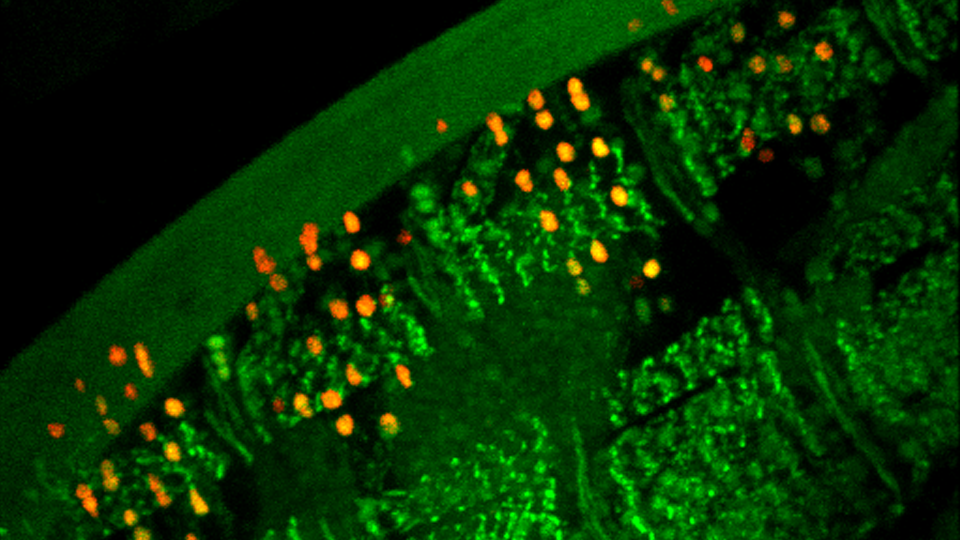
In this paper from von Diezmann and Rog in The Journal of Physical Chemistry, the authors established a method for using single‑molecule localization microscopy to resolve nanoscale dynamics within living tissue of extruded C. elegans gonad. Based on previous findings of preservation of motion in oocytes in extruded gonads within a nutrient‑rich environment, researchers adapted a similar methodology to examine meiotic functions. This process of sample preparation provided the necessary means for providing optimal optical properties and dye penetration into the sample while maintaining physiological conditions to allow for single‑molecule tracking experiments. Using extruded gonads resulted in reduced auto‑fluorescence and increased exposure of the tissue to surrounding buffer, enabling the use of the genetically encoded enzyme, HaloTag, and its corresponding HaloTag ligand coupled to the superior live‑cell single‑molecule imaging probe, PAJF549. In comparison to the photoconvertible protein mMaple3, PAJF549 offered greater photostability and higher photon output resulting in longer, more robust tracking. Bruker’s Vutara 352 super-resolution microscope was used to acquire single-molecule tracking data that suggest axis proteins fall into two categories, statically bound to chromatin or freely diffusing in the nucleoplasm. Furthermore, the diffusive proteins appear to selectively explore chromatin‑rich regions, avoiding the nucleolus. By combining well-established gonad extrusion protocols with improved single‑molecule tracking strategies, researchers have established a valuable tool for investigating chromosome organization and studying the molecular dynamics associated with this process.

      KEY TERMS:

  • Single-molecule localization microscopy, Multi-modal investigation, Meiosis, Cell division, Chromosome organization