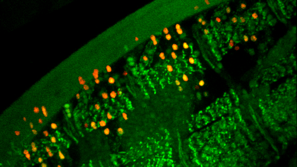
The nervous system is formed through cell cell connections, at specialized contact sites called synapses, forming neuronal circuits. How the nervous system correctly builds these connections has remained a mystery, but synaptic adhesion proteins, proteins expressed both pre- and postsynaptically, have been proposed to determine the specificity of connections. In the paper from Kurshan et al. the authors present data that a short form of the synaptic adhesion protein neurexin is required for assembly of presynaptic components even though this isoform lacks the canonical extracellular adhesion domains. Suggesting, that it is not its cell adhesion properties, but a direct function of this protein. They also discovered that neurexin worked in parallel with the Wnt receptor, Frizzled, but that this function was independent of its ligand Wnt and in fact Wnt resulted in reduction in synapses through removal of the Frizzled receptor from the plasma membrane by endocytosis. Together these results show how positive and negative synaptic development factors come together to help wire the nervous system.
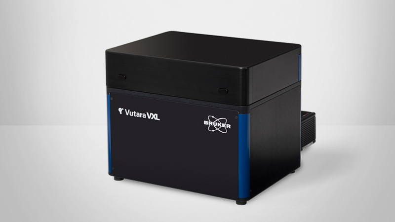
The authors use a combination of genetics, confocal microscopy, electrophysiology and superresolution mcroscopy with the Vutara 352; in Caenorhabditis elegans to reveal that a short form of neurexin is required for proper development of synapses. While confocal microscopy was useful in determining whether synapses were present or absent from the nervous system, super resolution microscopy was required to determine the substructure of the synapse in these mutants. The authors performed live super resolution microscopy using the Vutara 352 on C. elegans expressing Skylan-S and Halo-tagged synaptic proteins labeled with the Janelia fluor JF-646 (available for free to academic labs from Luke Lavis at Janelia, HHMI). The authors relied upon the 3D multi-color imaging to be able to image the synapses in a whole animal several microns from the coverslip. Confocal microscopy was not sufficient for this experiment because the average C. elegans synapse is approximately 400 nm across, while the active zone is on the order of 100-200 nms, below the diffraction limit of light. Using the Fourier Ring Correlation program included in the advanced statistics package in the Vutara SRX software the authors were able to determine the overall resolution of their images to be 40 nm laterally and 70 nm axially. Further, by using the DBSCAN algorithm, also included in the SRX software, the authors were able to determine the degree of clustering of each of the synaptic proteins tested. This analysis revealed that neurexin colocalized with calcium channels at the center of the synapse (the active zone, where the signal is propagated between cells), and this colocalization was higher than that of other active zone components suggesting there may be a functional connection between calcium channels and neurexin. Further, in neurexin mutants calcium channels were greatly reduced at the active zone, in both number and in the size of the calcium channel clusters present suggesting that neurexin is required for proper calcium channel clustering at the synapse. This work could only have been accomplished using he 3D capabilities of the Vutara 352 super resolution microscope along with the advanced statistics package included in the Vutara SRX software.