
Imaging Applications in Neuroscience: Assessing Neuronal Structure and Function
Explore Bruker's innovative solutions for seeing the biology of life.
This 2-hour, four-part virtual workshop features several brief technical presentations, each exploring a different fluorescence microscopy system. Key topics include:
- Executing holographic optogenetics experiments with a multiphoton microscope
- Imaging large cleared biological samples with light-sheet microscopy
- Utilizing single-molecule localization techniques to advance neuroscience research
- Performing precise spatial multiplexing in brain tissue
Contact us
For more information about this event or the products and techniques featured in it, please contact us. Follow @BrukerFM on Twitter for event, product, and webinar updates.
Featured Presentations & Technical Demonstrations
Ewa Zarnowska, Ph.D., Sales Applications Scientist, Bruker
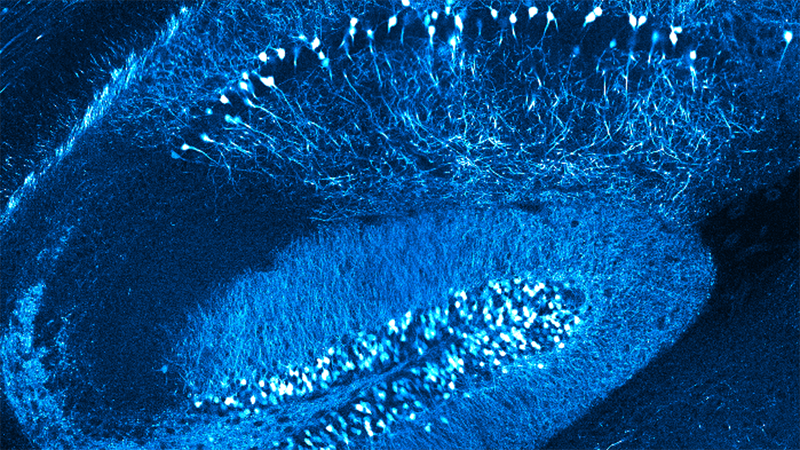
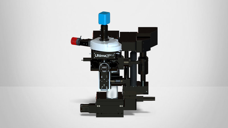
Optogenetics is an ingenious research technique that can help to solve the mystery of the brain’s works. In the process, the neuronal cells are virally transduced to be receptive to the light which is then used for manipulation of brain cells such as to activate them or to inhibit them.
Brain tissue is highly photon scattering where networks of neuronal cells are intricate and span across 3-dimensions. Therefore, observation and optical manipulation of the brain’s cells require advanced technical developments that provide adequate time- and spatial-resolutions.
Bruker’s answer to the needs of modern neuroscience was to design the state-of-the-art all-optical multiphoton workstation, the Ultima 2Pplus, which has which has larger than others field of view and is equipped with...
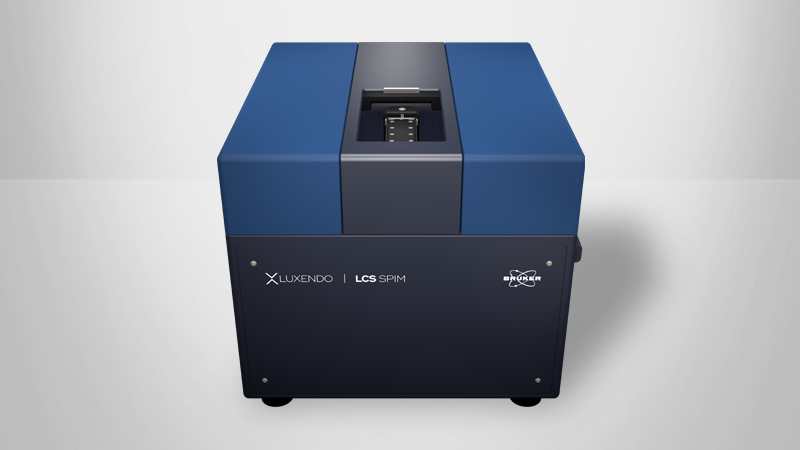
Jürgen Mayer, Ph.D., Senior Application Specialist and Product Manager, Luxendo
Imaging volumetric biological tissue with cellular or sub-cellular resolution has become a key factor in neuroscientific research. Light-sheet microscopy is the state-of-the-art methodology to achieve this, enabling non-destructive data acquisition of intact organs such as an entire mouse brain.
A prerequisite for optical imaging in large (i.e. mesoscopic) samples is tissue clearing. There are numerous different clearing techniques, but all of them have in common that they render biological samples optically transparent. The combination of cleared tissues with light-sheet microscopy is an ideal synergy that allows addressing new questions in neurobiology.
Minimizing sample mounting time, and a fast acquisition followed by a robust processing pipeline is important, especially as...
Winfried Wiegraebe, Ph.D., Product Manager for Super-Resolution Microscopy, Bruker
Lauren Gagnon, Ph.D., Appliations Scientist, Bruker
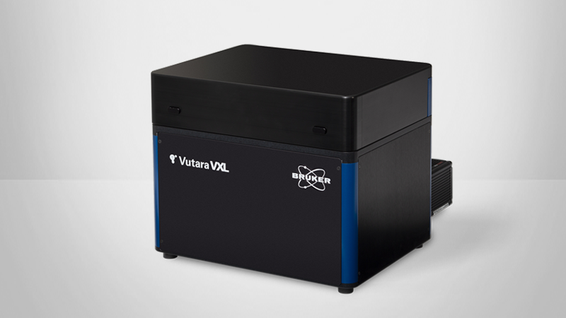
Single-Molecule Localization Microscopy (SMLM) was essential in tackling neuroscience questions about the organization of synapsis and the internal structure of neurons. With an optical resolution of 20 nm, SMLM is uniquely qualified to address biological mysteries that require specific labeling as used in fluorescence microscopy but higher resolution than can be achieved with diffraction-limited microscopy.
In addition to applications, we will discuss the basics of super-resolution microscopy and different implementations like dSTORM, PALM, and DNA-PAINT. We will discuss how the unique features of the Bruker Vutara VXL – its workflow-oriented software, biplane detection for improved z-resolution, and fluidics for multiplexed applications – will help you focus on neuroscience and getting superior results.
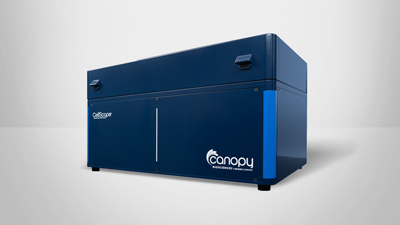
Adam Northcutt, Ph.D., Senior Scientist, Canopy Biosciences
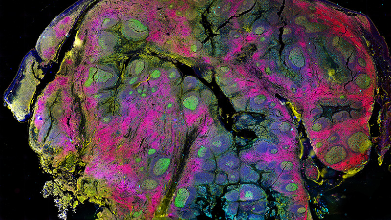
Understanding the spatial distribution of key neuronal cell populations is critical in advancing our understanding of disease to inform the development of novel therapeutics. Here we present the analysis of fresh frozen (FF) tissue samples from mouse brain using a novel precise spatial multiplexing technology called ChipCytometry, which combines iterative rounds of targeted fluorescent staining with high dynamic range imaging to facilitate quantitative phenotyping with single-cell resolution. Standard FCS files are generated from multichannel OME-TIFF images, enabling identification of cellular phenotypes via flow cytometry-like hierarchical gating. In this study, a 13-plex assay was used to identify and quantify relevant cellular phenotypes and subtypes for neurobiology and neuro-oncology applications. The results show...
Speakers
With introduction, live Q&A, and closing remarks led by:
Philip Golding, Fluorescence Microscopy Sales Manager Sales Manager, Bruker

Ewa Zarnowska, Ph.D.
Sales Applications Scientist, BrukerEwa is a neuroscientist who specializes in advanced microscopy and electrophysiological techniques. She received her Ph.D. degree in Biophysics from the Medical University of Wroclaw, Poland. She was a tenured Assistant Scientist in the Department of Anesthesiology at the University of Wisconsin in Madison.

Jürgen Mayer, Ph.D.
Senior Application Specialist and Product Manager, LuxendoDr. Jürgen Mayer is a Senior Application Specialist and the Product Manager for cleared system microscopes at Luxendo, the light-sheet microscopy branch of Bruker’s FM division. Before joining Luxendo, during his pre- and post-doctoral research, he developed and implemented a method to compensate attenuation artefacts in light-sheet microscopy via multimodal imaging combining light-sheet with optical projection tomography.

Winfried Wiegraebe, Ph.D.
Product Manager for Super-Resolution Microscopy, BrukerWinfried Wiegraebe is the product manager for super-resolution microscopy at Bruker. He has close to 30 years of experience in advanced microscopy in biology - including AFM, FCS, confocal microscopy, and super-resolution microscopy. Before joining Bruker, Winfried managed the Stowers Institute for Medical Research microscopy infrastructure in Kansas City, MO, US, and built the imaging pipeline at the Allen Institute for Cell Science in Seattle, WA, US. He studied Physics at the Technical University in Munich, Germany, and did his Ph.D. studies at the Max-Planck Institute in Martinsried, Germany.

Dr. Lauren Gagnon
Vutara Applications Scientist, Bruker
Dr. Lauren Gagnon graduated from Emmanuel College with a degree in Chemistry before completing her PhD in Chemistry at the University of Washington. Her thesis research focused on the development of novel approaches to single molecule localization microscopy using DNA barcoded antibodies.

Adam Northcutt, Ph.D.
Senior Scientist, Canopy BiosciencesAdam Northcutt is a Senior Scientist at Canopy Biosciences focused on application development for the ChipCytometry platform. Adam holds a Ph.D. in Molecular Biology from the University of Missouri with an emphasis in neuroscience and bioinformatics.