Model Systems to Trace Cancer Evolution, Treatment and Emergence of Residual Disease
See how organoid models can be used to study cancer recurrence
In this presentation, Dr. Martin Jechlinger (MOLIT Institut gGmbH; EMBL) discusses his research into why cancers reoccur after apparently successful treatment. Using organoid models that recapitulate tumor progression, regression, dormancy, and relapse, he investigates how residual cancer cells survive therapy and what drives their eventual re-emergence as recurrent disease.
Topics covered include:
- Modeling tumor regression, residual disease, and relapse in organoid systems
- Using live imaging to track clonal behavior and tumor cell fate over time
- Identifying the metabolic and epigenetic features associated with cancer recurrence
Model Systems to Trace Cancer Evolution, Treatment and Emergence of Residual Disease
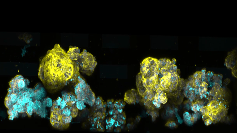
In this age of precision medicine, mortality due to breast cancer is primarily due to tumor relapse after initial targeted therapy. Cells left over from the primary tumor, remain dormant over time begetting the title of “residual disease”. These cells can give rise to refractory relapse tumors in patients many years (even decades) after primary tumor remission. Thus, there is an urgent need to fully understand the molecular basis of residual disease so we can target and limit this disease reservoir. However, the mechanism of establishment of residual disease remains elusive. This is primarily due to the lack of availability of patient samples during tumor regression. Our studies and efforts aim to better characterize residual disease using tractable models that faithfully recapitulates human disease. In conjunction with 3D organoid culture approaches, have been used to obtain a pure residual disease substrate, which adopts a deregulated metabolism. We further exploit this approach for live cell imaging at a single cell resolution using SPIM (Selective Plane Illumination Microscopy) to understand spatial and clonal dynamics of emerging and treatment refractory tumor cells.
Featured Technologies:
Contact:
Find out more about the technology featured in this webinar or our other solutions for light-sheet microscopy:
Speaker

Martin Jechlinger, Ph.D.
Head of VISION Laboratory at MOLIT Institut gGmbH;
Visiting Scientist at European Molecular Biology Laboratory (EMBL)