See What You Are Missing: Bruker Microscopes Fill the Gap Between Traditional Fluorescence and Electron Microscopy
The capabilities, resolution, and speed of conventional fluorescence and electron microscopes do not meet the needs of leading-edge life science research.
In this four-part webinar series, the Bruker applications team showcases our complete range of high-performance technology for the imaging and characterization of biological structures and processes. These featured technologies have capabilities that reach well beyond what is accessible with conventional methods and include:
Leading-Edge Instruments for Advanced Imaging and Characterization of Biological Materials and Processes
The imaging and characterization of biological structures and processes have moved beyond the capabilities, resolution and speed of conventional fluorescence and electron microscopes. For example:
- Eukaryotic cells consist of organelles and other subcellular structures so small they can only be resolved in detail with super-resolution methods;
- Such cells have membranes with mechanical properties best investigated at the nanoscale using atomic force microscopy;
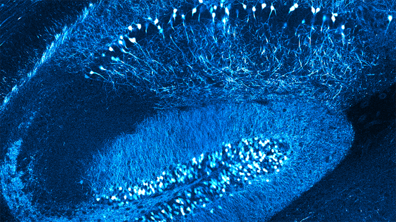
- Neurons form circuits in the brain so far from the surface only two-photon microscopy can reach them; and
- To visualize individual cells, neurons or even molecules in the context of an entire living organism requires light-sheet microscopy.
Bruker has developed a suite of advanced microscopes that collect information from cells, complex organ/tumor/tissue models or entire organisms in-vivo, information not accessible with traditional fluorescence or electron microscopy. In this four-part webinar, experts from Bruker discuss the technology behind atomic force, super-resolution, two-photon, and light-sheet microscopy, highlighting applications in which these technologies are essential to solving biological mysteries.
Find out more about the technology featured in this webinar or our other solutions for life science research:
Luxendo Light-Sheet Microscopes: Imaging Across Scales
Presenter: Lina Streich, Ph.D., Marketing and Applications Scientist, Bruker
Light-sheet microscopy is the coming-of-age gentle, high-resolution and fast, full 3D volume fluorescence imaging technique in fields as diverse as cell and developmental biology, neuroscience, oncology, plant research or biophysics.
With a strong focus on applications, Bruker Luxendo SPIM Light-Sheet configurations enable imaging of developing embryos or 2D and 3D culture systems like cell lines and organoids. This includes during multi-position experiments and under full environmental control which allows imaging over extended periods of time, even up to days. Furthermore, photomanipulation can be added to any of our SPIM system to support tissue photoablation, photobleaching, FRAP or optogenetic experiments.
In combination with tissue optical clearing techniques, Bruker Luxendo SPIMs allow in toto recording of very large specimens up to entire adult mice in a non-destructive fashion within minutes. Therefore, Luxendo SPIMs are ideal for studying the brain or the central nervous system, for analyzing organ development, or for investigating tumor structure and genesis in oncology.

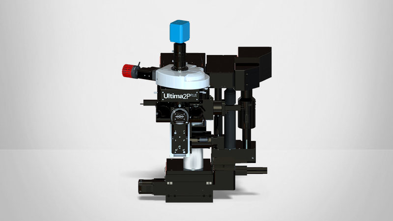
Ultima 2Pplus: Advances in Multiphoton Imaging and SLM Holographic Stimulation
Presenter: Romina Macco, Ph.D., EMEA Sales Applications and Customer Service Manager, Bruker
Multiphoton microscopy is a great approach for the investigation of cells, organoids, tissue slices, and organs in living animals with high penetration performance, better resolution along the Z dimension and limited sample heating and damage. This powerful technique is optimal to study the physiology and pathology of the brain in neuroscience, as well as to elucidate biological aspects of life sciences in a broad range of other biomedical applications including immunology and oncology.
Thanks to the high-level engineering design, the Bruker Ultima 2Pplus enables the imaging and investigation of a wider portion of biological samples. It also, offers a larger field of view, with respect to traditional multiphoton microscopes, without sacrificing signal uniformity, collection efficiency, resolution, and speed.
In addition to being a powerful imaging platform for both in vitro and in vivo studies, the Ultima 2Pplus is characterized by extraordinary photostimulation performances, thanks to the Spatial Light Modulator and the Electrically Tunable Lens modules. The combination of these two technologies results in fast 3D imaging and the holographic photoactivation of dozens of cells simultaneously and with high precision in the X, Y, and Z dimensions.
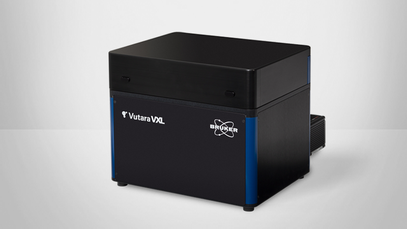
Single-Molecule Localization for Advanced Biological and Genomic Imaging: The Bruker Vutara VXL
Presenter: Winfried Wiegraebe, Ph.D., Vutara Super Resolution Microscopy Product Manager, Bruker
Most subcellular structures are smaller than 300 nm so traditional fluorescence microscopy does not have the specificity to resolve these small sizes. The single-molecule localization microscopy (SMLM) technology implemented in the Bruker Vutara VXL overcomes this limitation. With an optical lateral resolution of 20 nm, SMLM is uniquely qualified to address biological mysteries that require specific labeling as used in fluorescence microscopy, but with higher resolution than can be achieved with diffraction-limited microscopy. In addition to SMLM, the Vutara VXL implements imaging workflows for multiplexed chromatin tracing and smFISH applications. Thus. the system allows imaging, resolving, and quantifying cellular structures, molecular machines, proteins, RNA, and chromosomal structures.
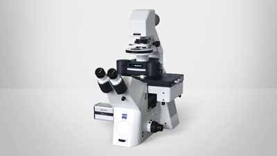
Atomic Force Microscopes for Imaging and Characterizing Biological Materials and Processes
Presenter: Torsten Mueller, Project Manager, Ph.D., Bruker
In the past decades, atomic force microscopy (AFM) has become standard in the high-resolution structural analysis of samples ranging from single molecules to complex macromolecular systems. Unlike other high-resolution imaging techniques, it does not require any specific sample modification (except for surface deposition) and therefore reduces the risk of introducing artifacts through sample preparation. In recent years, there has been an increased demand for the application of novel developments featuring high-speed AFM imaging (>10 frames/sec), quantitative nanomechanical characterization of samples, and advanced feedback imaging modes that tweak the maximum attainable resolution. Furthermore, the examination of specific molecules or features carrying immunochemical information has been made possible by combining AFM with recent developments in optical/fluorescence microscopy, therefore complementing the advantages of both techniques and enabling true correlative microscopy.