
How does XRF Work?
X-Ray Fluorescence (XRF) Spectrometry
The XRF technologies provide elemental analysis of a huge variety of materials including metals, alloys, polymers, ceramics, geological materials, petroleum products, soil, paint and much more. What is XRF? Which XRF techniques are available? What elements can be detected? How accurate and fast is the analysis? Find the right solution for your analytical needs.
The Principles of XRF Spectrometry
XRF describes the process where some high-energy radiation excites atoms by shooting out electrons from the innermost orbitals. When the atom relaxes, that is, when outer electrons fill inner shells, X-Ray fluorescence radiation is emitted. All this happens without touching or damaging the sample.
The emitted radiation is very much like a fingerprint of the atom. Copper fluorescence looks very different from zinc fluorescence and from fluorescence of any other element in the periodic table. That is why XRF is one of the most straightforward and convenient ways to do elemental analysis and is used for a vast number of industrial, research and educational applications. Here, the derived data can be used to obtain qualitative, semi-quantitative and quantitative information about the major, minor and even trace elements in a sample.
So, the question only is: What do you need XRF for? Will it be trace element analysis? Standardized high-precision quantitative analysis in a cement plant? 2D failure analysis and production control? Sorting metal in a scrap yard, or pre-screen rock formations at an excavation site?

The difference between EDXRF and WDXRF
Any XRF instrument nowadays comes with an X-ray tube for excitation of the atoms in the sample and a detector, for registering of the fluorescence radiation. The tubes can be water-cooled high-power tubes with an output of 4000 W, or tiny, thumb-sized 4 W tubes for mobile devices. On the photon detection side, there are basically two different technologies: Energy-Dispersive (EDXRF) or Wavelength-Dispersive (WDXRF). Even though the energy E of a photon and its wavelength λ are virtually exchangeable (because of the fixed relation E = (c ∙ h)/λ , with c being the speed of light and h th Planck constant) the way of sorting the photons with respect to either E or λ is quite different:
A wavelength-dispersive spectrometer treats the X-rays as waves and uses some regular structure (gratings or crystals) to cause interference patterns that allow for remarkably high spectral resolution. The energy-dispersive detectors treat the X-rays as particles. They work like throwing a bowling balls (the photons) into a ball pit (the detector) to see how many small plastic balls (electrons) are ejected by each impact. Heavier or faster bowling balls (higher energies) will cause more plastic balls to be ejected.
XRF Technologies
There are many techniques derived from XRF. Some are very sensitive (TXRF), others very precise (WDXRF). Some for valuable, delicate objects that must stay untouched (Micro-XRF/Macro-XRF), others rather for materials used for and derived from industrial processes (Direct & Polarized EDXRF). Some give you the exact concentrations of all elements contained in the sample (WDXRF); others can tell you exactly where some inclusion is in the sample (Micro-XRF). Most are laboratory instruments; some can be carried around in one hand (Handheld-XRF).
Some techniques are parts of ISO and ASTM standards in industrial quality management processes, the versatility of others makes them more and more frequent in R’n’D labs.
The selection of the optimal spectrometer is driven by the analytical requirements:
- Portable, benchtop, or lab-based element analysis
- Number of samples to measure per day
- Material/sample type and preparation
- Elements of interest, concentration ranges & limits of detection
- Accuracy and precision
- Sample structure and structure size
Specifications
| Technologies | Portable EDXRF | Benchtop EDXRF | Benchtop EDXRF | Benchtop EDXRF | Benchtop EDXRF | Benchtop WDXRF | Sequential WDXRF | Simultaneous WDXRF |
| Direct EDXRF | Polarized EDXRF | Micro-XRF | TXRF | |||||
| Solid samples | +++ | +++ | ++ | +++ | ++ | +++ | +++ | +++ |
| Liquid samples | + | +++ | +++ | ++ | +++ | +++ | +++ | - |
| Element range | (F)Mg-Am | (C)F - Am | Mg-Am | (C)Na-Am | (Na)Mg-Am | (C)F - Am | (Be)B - Am | (Be)B - Am |
| Mobility | +++ | + | ++ | ++ | (+)+ | + | - | - |
| Speed | +++ | ++ | ++ | ++ | ++ | ++ | ++ | +++ |
| Accuracy & precision | + | ++ | +++ | ++ | ++ | ++ | +++ | +++ |
| 2D spatial resolution | ++ | + | - | +++ | - | - | ++ | - |
| Concentration range | Ppm to wt.-% | Ppm to wt.-% | Sub-Ppm for S, Cl, P | Ppm to wt.-% | Ppb to wt.-% | Ppm to wt.-% | Sub-Ppm to wt.-% | Ppm to wt.-% |
| Read more | Portable XRF | Direct EDXRF | Polarized EDXRF | Micro-XRF | TXRF | Benchtop WDXRF | Sequential WDXRF | Simultaneous WDXRF |
Direct & Polarized EDXRF
Benchtop Energy-Dispersive X-Ray Fluorescence (EDXRF) spectrometers are compact in size and have a relatively simple setup. A 50-Watt X-ray tube is used to excite the sample and a Silicon-Drift-Detector (SDD) to count the number and energy of the characteristic X-ray photons. The SDD records the entire energy spectrum at once and thus multiple elements can be detected simultaneously. Direct EDXRF spectrometers are versatile devices, which make use of a closely coupled beam path to measure major, minor, and trace elements in all kinds of liquids, powders, grains, solids, and bulk samples. Polarized EDXRF spectrometers utilize monochromatic X-rays to enhance signal-to-noise ratio for elements like S, P, and Cl in petrochemicals, to achieve ultra-low detection limits even with a compact device.

Handheld / Portable XRF
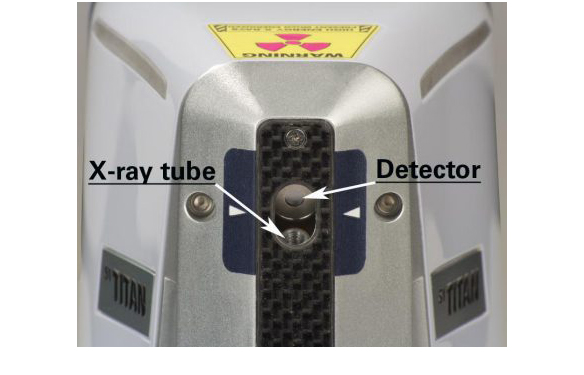
The smallest possible complete XRF spectrometers are small enough to run on batteries and be carried in one hand. Still, they can do a full positive material identification within seconds. A handheld-XRF has the same components as larger EDXRF instruments but packed into a much smaller volume. Despite of their size, these instruments are very accurate and sensitive for all elements starting from magnesium. That’s because the tube and the detector, naturally, are very close to the sample. So not only can they be carried around in a scrap yard to sort metals, but also can they be used in-field for trace element analysis in geo-exploration campaigns.
Micro-XRF
Micro-XRF is an energy-dispersive method which uses a polycapillary optic to guide the excitation X-rays into a micrometer-sized spot on the sample. Thus, it cannot only tell you what elements are in a sample and how much of them, but also where exactly they are. Especially when combined with a fast-scanning stage, this method is ideal for measuring and understanding inhomogeneous samples. With micro-XRF almost any kind of sample can be measured. Solids, powders, and liquids. The need for sample preparation is minimal and – like all XRF methods, the sample is not damaged in the process of measuring. This makes micro-XRF an ideal pre-screening technique, not only in forensics. Also, in geology this sort of material analysis is almost perfectly adapted to the analytical task.

TXRF

Yet another energy-dispersive technique, Total-reflection XRF (TXRF) is using every possible tweak to optimize the signal to noise ratio and, hence, the limits of detection. The specific difference to all other EDXRF techniques is the very shallow incident angle. It is indeed so shallow that the monochromatized excitation X-rays are totally reflected at a smooth substrate, hence the name. This geometry offers three main advantages:
- Without going into the substrate there is no scattered background signal from the substrate.
- By being reflected, the excitation beam goes twice through the sample which lies on that substrate.
- With the X-ray beam coming from one side and going to the other, the detector can be placed very close to the sample surface, without obstructing the beam. Thus, almost all the fluorescence produced in the sample is collected by the SDD. Minute amounts of sample material, down to microliters or micrograms, can be quantified and detection limits in the ppb-range are achievable with minimal sample preparation.
WDXRF
Wavelength Dispersive X-Ray Fluorescence (WDXRF) spectrometers utilize a more sophisticated setup when compared to EDXRF systems, enabling 1-2 orders of magnitude higher sensitivity. WDXRF systems are equipped with powerful X-ray tubes (up to 4.000 W), several optical components (e.g., filter, collimators) and make use of analyzer crystals to separate X-ray photons emitted by the samples based on their wavelength, following Bragg’s law. Sequential floor standing WDXRF and benchtop WDXRF systems spectrometers are equipped with a goniometer, which changes the angles and crystal type for each wavelength accordingly, i.e., the elemental concentrations are measured sequentially. Large simultaneous WDXRF spectrometers make use of fixed channels to measure multiple elements at once and achieve shortest measurement times.

Typical XRF Applications
XRF technology can be found in countless environments where elemental analysis is essential. Process monitoring and quality control in mining, cement & building materials, petrochemicals & polymers, food & animal feed, and metal production are just some examples. Portable devices are particularly useful during field exploration campaigns and for scrap metal sorting. XRF is also frequently applied in art, conservation & archaeology and in forensics. The versatility and the simple sample preparation make XRF an inevitable tool for many service labs and governmental institutions as well as in academia and research.
Read more about Bruker's solutions for:
- Materials Science Research
- Minerals, Mining & Petrochemical
- Industrial
- Semiconductor & Nanotech
- Food Analysis & Agriculture
- Environmental & Forensics