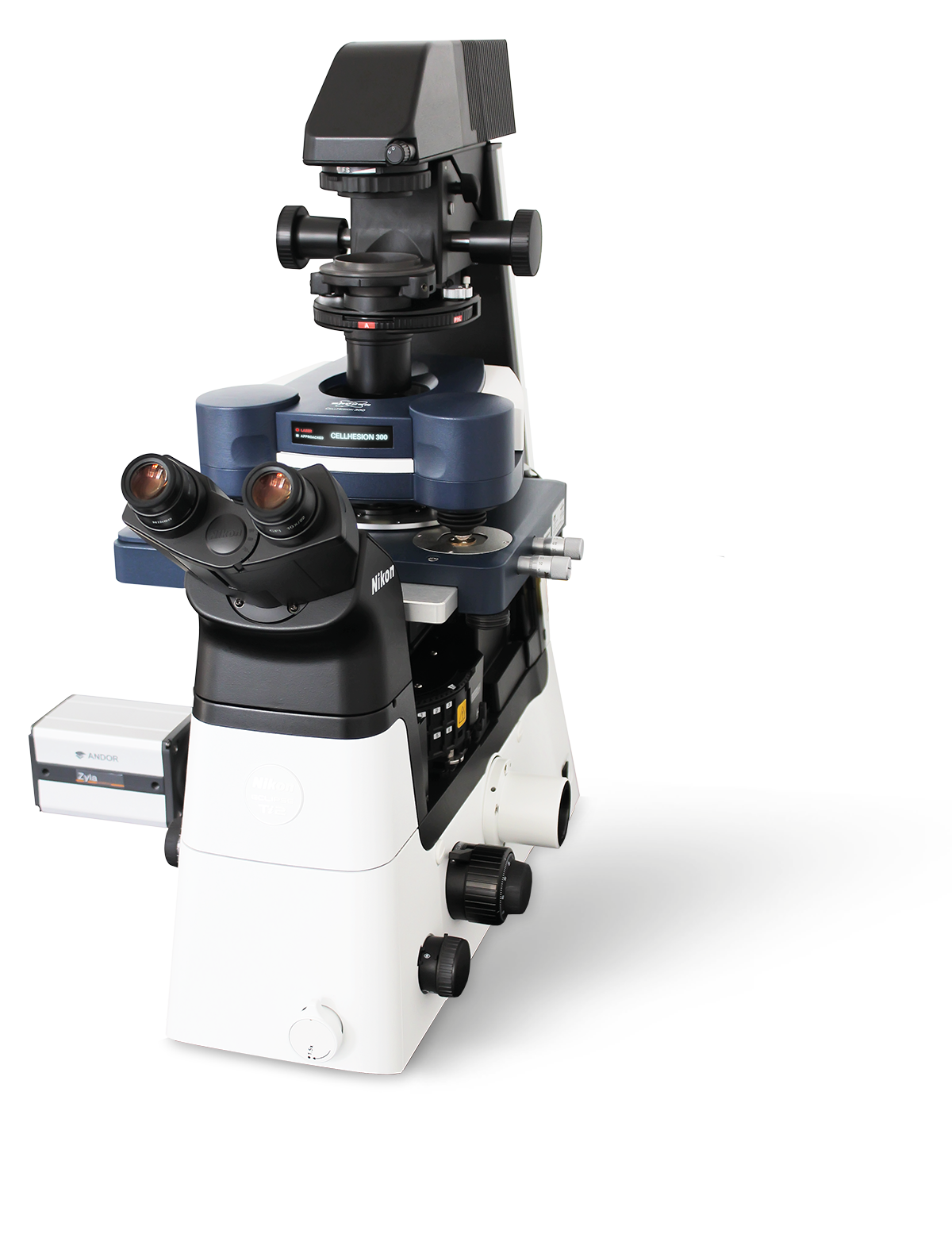
Cell Hesion 300
CellHesion® 300
いくつかの機能が自動化されたCellHesion®300は、細胞-細胞、細胞-組織、細胞-基板間の相互作用を1分子レベルで高感度に測定するための理想的なツールです。その高感度測定により、生きた生体システムの構造、形態、ナノ力学的特性を迅速かつ容易に取得でき、さまざまな病理疾患におけるそれらが果たす役割について重要な知見が得られます。この革新的なシステムは、生物物理学、生化学、インプラント研究、創傷治癒、発達生物学、幹細胞研究、感染症生物学および免疫反応研究等のアプリケーションに対して新たな可能性をもたらすでしょう。
Understanding Biomechanics Made Easy
CellHesion300は、高品質で再現性のある定量データを提供します。そして、高度な自動化による、スループットの向上、生物医学および臨床研究に必要な生産性、パフォーマンス、および統計的有意性を実現します。最大接着力、個々の解離する力、細胞間相互作用などの重要なパラメータが自動的に革新的なソフトウェア ソリューションによって各データセットから決定されます。
CellHesion300が、どのように構造と機能の関係や、細胞や組織に対する機械的な力の影響を研究し、発生生物学、組織工学、ナノメディシンなどの分野への応用の新たな可能性を開くために使用できるかを、短いビデオでご覧ください。
Only CellHesion 300 delivers:
- 測定の自動化によるデータスループットの最大化
- 生理的条件下における生体試料のナノ力学計測
- 組織生検に最適な、広いサンプルエリアでの迅速かつシンプルな測定領域選択。
- 視覚的にわかりやすいソフトウェア
イメージ(右):単一細胞を用いた相互作用を検出する実験では、単一の生細胞を生化学的にプローブに結合させます。例えば、カンチレバーの表面修飾を介して)細胞を結合標的と接触させ、設定した力を細胞に加えます。( 1 )
ユーザーが設定した結合時間経過後、プローブを引き離すことにより、標的と接触した細胞をターゲットから分離します。( 2 )
引き剥がす力などは、カンチレバーのたわみの定量化によって解析されます。
革新的なマルチパラメトリックナノメカニカルマッピング
視覚的にわかりやすいソフトウェア、直感的なユーザーガイダンス、および自動化された検出システムによる非常に使いやすい操作性と迅速なデータ取得。
測定用ソフトウェアでは使いやすいスクリプトツールが、解析ソフトウェアでは、強力なバッチデータ処理機能による大規模データの分析と定量化が可能になります。
実験の拡張性
- 標準の100 μm Zスキャナに加え、オプションの15 μm Zスキャナの2つのZスキャナを連動させたNestedScanner機能により、大きな高低差のある試料に対しても高速性を備えた粘弾性評価が可能になります。
- 生きている細胞等を生理的に近い状態で研究するための豊富なアクセサリー(試料温度やCO2濃度など環境条件のコントロール)。
Automate Investigation of Large Samples
ユーザーは光学顕微鏡画像からフリーハンドで選択した任意の2 D 形状でフォースマッピング計測を行うことができます。(右上図参照)光学画像を連結させた光学タイリングを使用すると、非常に大きいタイリング画像領域から関心のある複数の領域を事前に選択することにより、それらの領域の自動測定が簡単かつ効率的に実行できます。これらは改良された電動ステージの精度により、これまで以上の精度と速度が実現できました。
光学顕微鏡とシームレスに統合
CellHesion 300 は、高度な超解像能力を備えた最新の光学顕微鏡にシームレスに統合できるため、生きた生物学的サンプルの包括的な特性評価に向けた光学画像とAFMデータの相関データセットの提供が可能です。また、CellHesion 300 は、粗い表面、密集した細胞層、および非常に皺の多い組織サンプルのバイオメカニカル研究に最適なソリューションです。
サンプル提供: Prof. Dr. Ansgar Petersen, BIH, Center for Regenerative Therapies, Charité Medical University, Berlin, Germany
Selection of Scientific Publications Using the CellHesion Technology
- Abuhattum et al., Adipose cells and tissues soften with lipid accumulation while in diabetes adipose tissue stiffens. Sci Rep 12, 10325 (2022).
- Michael et al., Measuring the elastic modulus of soft culture surfaces and three-dimensional hydrogels using atomic force microscopy. Nat Protoc 16, 2418–2449 (2021).
- Liebsch et al., Quantification of heparin’s antimetastatic effect by single-cell force spectroscopy. J Mol Recognit. 34, e2854 (2021).
- Möllmert et al., Zebrafish Spinal Cord Repair Is Accompanied by Transient Tissue Stiffening. Biophys J. 118(2), 448-463 (2020).
- Shen et al., Reduction of Liver Metastasis Stiffness Improves Response to Bevacizumab in Metastatic Colorectal Cancer. Cancer Cell 37(6), 800-817 (2020).
- Rheinlaender et al., Cortical cell stiffness is independent of substrate mechanics, Nat. Mater. 19, 1019–1025 (2020).
- Aaron at al., Quantification of heparin's antimetastatic effect by single-cell force spectroscopy, J Mol Recognit., 1–11 (2020).
- Krieg et al., Atomic force microscopy- based Mechanobiology, Nature Reviews Physics 1, 41–57 (2019)
- Stylianou et al., Review Article: Atomic Force Microscopy on Biological Materials Related to Pathological Conditions, Andreas, Scanning, 8452851 (2019)
- Thompson et al., Rapid changes in tissue mechanics regulate cell behaviour in the developing embryonic brain. eLife 8:e39356 (2019)
- Miroshnikova et al., Adhesion forces and cortical tension couple cell proliferation and differentiation to drive epidermal stratification, Nat Cell Biol 20, 69–80 (2018)
- Elias et al., Tissue stiffening coordinates morphogenesis by triggering collective cell migration in vivo, Nature 554, 523-527 (2018)
- Bharadwaj et al., aV-class integrins exert dual roles on a5b1 integrins to strengthen adhesion to fibronectin, Nature Communications 8, 14348, 1-10 (2017)
- Friedrichs et al., A practical guide to quantify cell adhesion using single-cell force spectroscopy. Methods (2013)
CellHesion Data Gallery
Bruker’s BioAFMs allow life science and biophysics researchers to further their investigations in the fields of cell mechanics and adhesion, mechanobiology, cell-cell and cell-surface interactions, cell dynamics, and cell morphology. We have collected a gallery of images demonstrating a few of these applications.
The Widest Range of Accessories in the Market
Optical systems/accessories, electrochemistry solutions, electrical sample characterization, environmental control options, software modules, temperature control, acoustic and vibration isolation solutions and more. Bruker provides you with the right accessories to control your sample conditions and to perform successful experiments.
Accessories to extend your BioAFM capabilities
Bruker offers an extensive range of BioAFM system add-ons, accessories, and modes to deliver maximum experimental and sample control, superior versatility, and enhanced useability. These options extend the range of applications and experiments supported by Bruker BioAFMs far beyond what is possible with any other BioAFM system on the market today.
Available options include optical systems/accessories, electrochemistry solutions, electrical sample characterization, environmental control options, software modules, temperature control, acoustic and vibration isolation solutions, and more.
Browse our online accessories database or download our accessories handbook to learn more.
Watch Recent BioAFM Webinars
Our webinars cover best practices, introduce new products, provide quick solutions to tricky questions, and offer ideas for new applications, modes, or techniques.
お客様の声
"非常に大きな高低差のある試料に対しても、CellHesion 300は対応可能であるため、臨床における組織の特性評価に極めて役に立ちます。"
Prof. Dr. Ansgar Petersen
BIH, Center for Regenerative Therapies
Charité Medical University, Berlin, Germany


