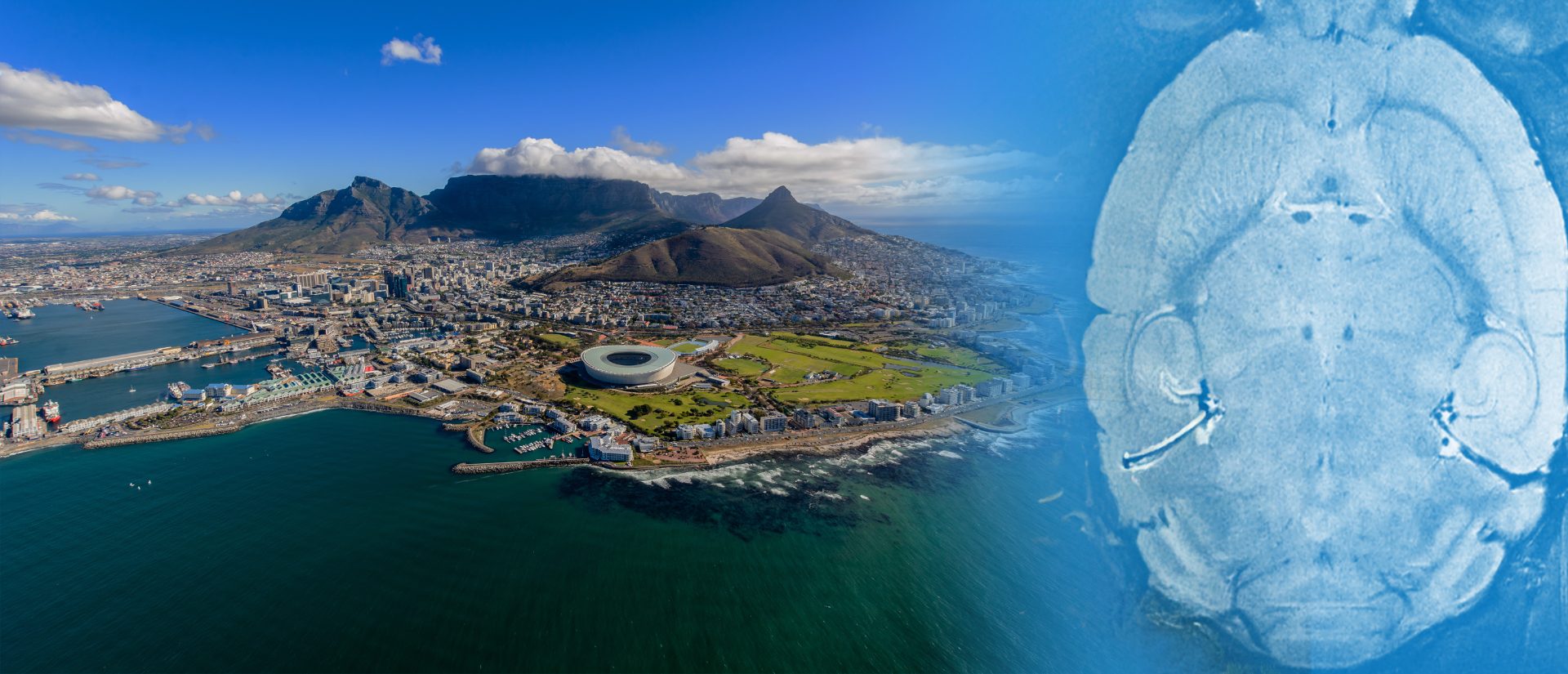
Bruker at ISMRM 2026
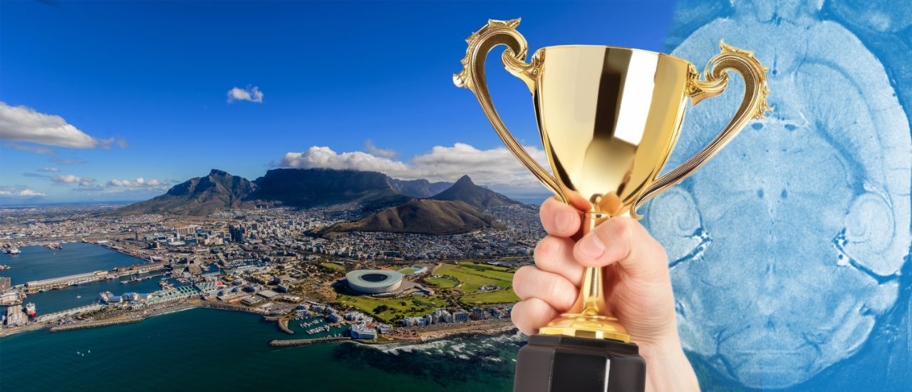
Bruker Preclinical Imaging is heading to Cape Town for ISMRM 2026 and we’re bringing a fresh way to navigate the “field”: the terrain of South Africa, the magnetic field, and your field of study.
Let Bruker Be Your Field Guide to Discovery as you explore our comprehensive portfolio—featuring UHF MRI for ultrahigh sensitivity and microstructural detail, BioSpec Maxwell for robust performance and streamlined workflows, Dynamis DNP for rapid, hyperpolarized metabolic imaging, PET/MR for advanced dual-modality insights, and ParaVision 360 for versatile, consistent, and reliable software solutions. Whether your focus is oncology, neurology, or metabolic imaging, Bruker’s technologies are designed to empower your research and accelerate your path to new breakthroughs.
At the Bruker booth, you’ll find hands‑on demos, expert consultations, and practical workflows that move science from idea to insight—faster. Come meet our applications scientists, swap best practices with our community, and leave with tangible ways to elevate sensitivity, throughput, and decision‑making in your lab.
We look forward to seeing you in Cape Town, 9–14 May 2026.
Bruker Scientific Workshop at the ISMRM
Saturday May 9th, 2026
Westin Cape Town (connected to CTICC)
The Grand Ballroom – Old Harbour Level
Registration opens at 12:30 with light lunch provided
First talk will start at 13:00
Agenda
| Time | Topic | Speaker |
| 13:00 | Introduction and Welcome from Bruker | Tim Wokrina & Guillaume Tetard |
| New Metabolic Perspectives along the Path to Translation | ||
| 13:20 | Keynote - Preclinical Metabolic Imaging | Anke Henning |
| 14:05 | Translational Molecular MRI with CEST: From Preclinical UHF Imaging to Clinical Applications | Philip Boyd |
| Coffee Break | ||
| 14:50 | Bruker Preclinical Imaging News and Highlights | Tim Wokrina |
| 15:40 | Bruker MRI Award Presentations | Tim Workrina & Winners |
| Coffee Break | ||
| Tracing Disrupted Pathways | ||
| 16:35 | Engineering Metabolic MRI: Technology Development, Disease Translation, and Cross-Polarization Innovation | Renuka Sriram |
| 17:00 | Blood Brain Barrier assessment with MR-PET | Gary Cowin |
| 17:25 | Closing Remarks | Tim Wokrina & Guillaume Tetard |
| 17:30 – 17:45 | Transportation to Bruker Night | |
Bruker Night
Saturday May 9th at 18:00
Cabo Beach Club, V&A Waterfront
12 S Arm Rd, Victoria & Alfred Waterfront, Cape Town, 8002, South Africa
Transportation will be provided to and from the CTICC to the Cabo Beach Club.
Bruker is proud to host our guest speakers for this year’s Scientific Workshop.

Anke Henning, Ph.D.
An expert in medical imaging technology development, Dr. Henning has developed novel magnetic resonance imaging methods for noninvasive visualization of disease-related structural, functional, and metabolic changes in the human brain, spinal cord, and heart.
Her expertise is in ultra-high field magnetic imaging technology, magnetic resonance spectroscopy, and non-proton imaging. She also directs clinical research with respect to brain imaging in psychiatric disorders, spinal cord imaging in traumatic injury, and imaging of the human heart. Dr. Henning has expanded her research interests as a Cancer Prevention and Research Institute of Texas Scholar in Cancer Research to include imaging glioblastoma with the ultimate goal of enhancing the treatment of this aggressive, incurable brain cancer.
Preclinical Metabolic Imaging
Magnetic Resonance Imaging (MRI) and Spectroscopy (MRS) offer powerful tools to probe metabolism. By leveraging advanced techniques like high resolution 1H, 31P and 2H MRSI, incorporating stable isotopes such as 13C and 2H, hyperpolarization, chemical exchange saturation transfer (CEST), and multimodal PET-MRI, we can gain deep insights into metabolic processes relevant to basic research as well as clinical-translational science. Importantly, many of these approaches are not limited to preclinical studies—they hold strong potential for translation to human applications, enabling visualization and quantification of disease-related changes in human tissue. Such capabilities provide a foundation for mechanistic understanding of disease and for developing diagnostic and therapeutic strategies.
Philip S. Boyd Ph.D.
Dr. Philip S. Boyd holds a Ph.D. in Physics from Heidelberg University and leads the CEST Imaging Group at the German Cancer Research Center (DKFZ) in Heidelberg. His research focuses on developing and refining advanced CEST imaging techniques, including pH imaging and fat artifact correction. He combines experimental imaging, mathematical modeling, and data analysis to drive CEST applications from preclinical research to clinical translation.
Translational Molecular MRI with CEST: From Preclinical UHF Imaging to Clinical Applications
This talk presents translational molecular MRI research at the German Cancer Research Center (DKFZ) using Chemical Exchange Saturation Transfer (CEST), spanning preclinical ultra-high-field (UHF) imaging and applications at clinical field strengths. Preclinical studies at ultra-high magnetic fields—such as those performed on the DKFZ’s 9.4T Bruker system—play a key role in methodological development, enabling a deeper understanding of complex CEST signal behavior in vivo and paving the way for clinical translation. Building on these advances, applications in cancer and neuroimaging will be highlighted, including human studies at 7T and 3T. Complementary 31P MR spectroscopic imaging for non-invasive pH mapping in humans at 7T will also be presented.


Dr. Renuka Sriram
Dr. Renuka Sriram is an Associate Professor (in-residence) in the Department of Radiology and Biomedical Imaging at the University of California, San Francisco (UCSF). She serves as the Director of the UCSF Pre-Clinical Magnetic Resonance Imaging and Spectroscopy Core and is Co-Leader of Technology Research and Development (TR&D2) at the P41-funded Hyperpolarized Magnetic Resonance Technology Resource Center (HMTRC) at UCSF. She has co-authored over 60 peer-reviewed publications and serves as Principal Investigator on multiple federally funded grants.
Dr. Sriram’s research focuses on leveraging metabolic dysregulation in cancer and other pathologies to develop advanced magnetic resonance–based imaging biomarkers for disease detection, characterization, progression monitoring, and therapeutic response assessment. A central emphasis of her work is the application of dissolution dynamic nuclear polarization (dDNP) and hyperpolarized 13C magnetic resonance imaging for translational cancer research, particularly in urologic oncology.
Her program integrates innovative imaging technology development with biologically relevant model systems to accelerate clinical translation. She is actively developing biomarkers of tumor presence, aggressiveness, and early therapeutic response. In parallel, she is establishing a comprehensive compendium of clinically relevant preclinical models optimized for metabolic imaging. These platforms span ex vivo cell and tissue slice cultures adapted to physiologically relevant, MR-compatible bioreactor systems, as well as in vivo transgenic and orthotopic murine models utilizing patient-derived living tissues. Through these efforts, her work aims to bridge mechanistic metabolic insights with robust clinical implementation of hyperpolarized MR technologies.
Engineering Metabolic MRI: Technology Development, Disease Translation, and Cross-Polarization Innovation
Hyperpolarized ¹³C MRI has rapidly evolved from an experimental research platform into a clinically translated technique for probing in vivo metabolism in cancer and other diseases. This lecture will examine the key technical barriers that shaped this progression and the engineering innovations that enabled application-driven translation. Achieving this required the development of reliable polarization workflows, metabolite-selective RF pulse design, rapid spectroscopic imaging strategies, integrated engineering solutions—including bioreactor systems—and rigorous quantitative kinetic modeling to measure downstream metabolism of hyperpolarized substrates for treatment response assessment and biological insight. More recently, in collaboration with Bruker, our efforts have focused on advancing the underlying MR physics through the adaptation of cross-polarization methodologies to accelerate polarization buildup and enhance sensitivity. Collectively, these advances demonstrate how sustained, application-informed technical innovation—spanning urologic oncology, infection, and inflammatory disease—has established hyperpolarized ¹³C MRI as a versatile metabolic imaging platform while opening new opportunities at the interface of translational imaging and MR physics.
Gary Conwin
Associate Professor Gary Cowin is an Australian National Imaging Facility (NIF) fellow within the Australian Institute for Bioengineering and Nanotechnology at the University of Queensland. He has >30 years of clinical and preclinical research experience in MRI, PET, and CT. He is a specialist for imaging technology for researchers and commercial partners to maximise the use of new multimodal imaging technologies into their research.
Cowin is a leading scientist developing experimental, imaging, and analysis techniques to utilise the potential for simultaneously acquired MRI and PET data. This has focused on quantification of the blood-brain barrier integrity during uptake studies of new PET tracers. This allows gadolinium enhanced MRI gadolinium to independent assessment of vascularity and blood-brain barrier permeability during the delivery of the PET tracer. These new methods have been applied rodent models of traumatic brain injury, epilepsy, focused ultrasound opening of the blood-brain barrier, and Parkinson's disease. He also applies imaging expertise to agricultural and industrial programs
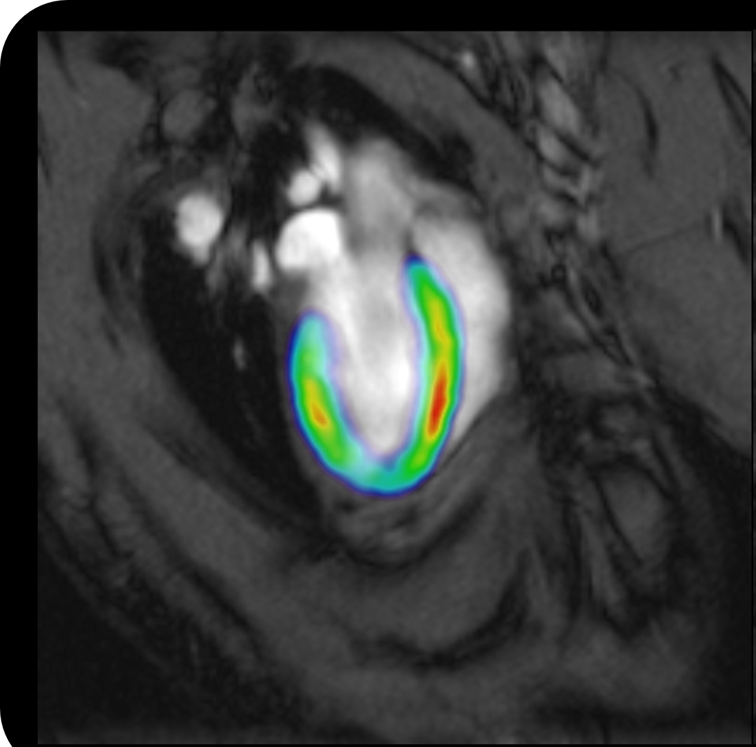
Blood Brain Barrier assessment with MR-PET
The blood brain barrier (BBB) is a unique challenge for designing and delivering new diagnostics and therapeutics. Simultaneous MR-PET enables independent assessment of the BBB permeability parallel to the delivery of a PET tracer. Mixing gadolinium contrast agents with the PET tracer in a single syringe allows simultaneous dynamic MR and PET acquisitions. Post processing of the dynamic MR data enables assessment of vascular delivery and BBB permeability. This will be demonstrated using transient opening of the BBB with focused ultrasound and in brain tumours.

Our Highlights

BioSpec Maxwell
Intelligent MRI in a small footprint

BioSpec Ultra-High Field MRI
The Ultimate in Preclinical MRI

Coils and Cradles
Perfectly Matched to your applications

ParaVision 360

Preclinical Imaging Community
Join our community of preclinical imaging users

Preclinical Imaging Drives Neuroscience
Improving knowledge of brain development, structure, and function