ParaVision 360
ParaVision 360 V3.7 available now!
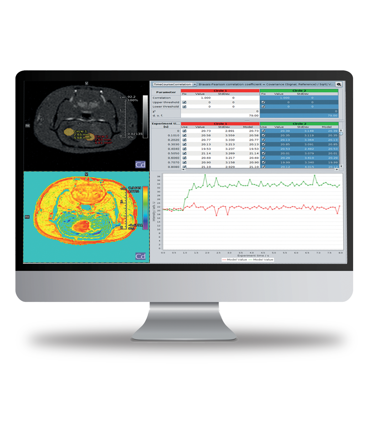
Focusing on consistent quantification, ParaVision 360 makes scanning even more intuitive, while maintaining the full flexibility that Bruker users praise. The enlarged MRI sequence portfolio, which is exclusive to Bruker, and the powerful image data evaluation functions lead to maximum proficiency in scanning and analysis, while the simplicity and uniformity across the range of instrumentation allows operators to put their focus on their research.
Highlights from the newest V3.7 release
B0 Distortion Correction for EPI
- Robust field map-based correction of geometric distortions
- Improved anatomical accuracy for key applications, e.g. fMRI and DTI
- Integration into the standard reconstruction pipeline
Enhanced Smart Noise Reduction
- Improved image quality with new „Crisp“ network
- Reduced flickering and improved image contrast with Image Series Denoising
- Significantly reduced reconstruction times
Improved PET studies
- Comprehensive isotope database for simple selection of most common isotopes
- Bowsher Prior reconstruction leveraging anatomical MRI to improve the PET image
- Expanded suite of color lookup tables including the Cold color palette for enhanced interpretability
Highlights from previous versions of ParaVision 360
Cross-modality Highlights
- Common terminology between instruments, common integrated multimodal workflows, and common image processing, viewing, and analysis tools and functionalities for optimal clarity
- Automatic 3D image fusion
- Drag and drop image processing
- Data browser with simple search functions for accessing stored protocols and recorded data
- Database and archiving and report creation
Highlights for MRI
- Magnet status display in ParaVision and remote instrument monitoring option
- Fast online decisions and real-time calculations on all channels
- MRI pulse program display
- Wobble control on magnet front touchscreen display²
- SLASER single voxel spectroscopy that is robust against B1 inhomogeneities and provides less chemical shift displacement
²For instruments with Animal Transport System
Highlights for PET
- Extensive reconstruction algorithms (e.g. MLEM, OSEM, MAP, FBP)
- MRI or CT based attenuation corrections with automatic loading of animal cradle and MRI coil and individual calculation of subject
- Wireless cardiac PET/MR imaging
- Scatter, randoms, isotope decay, and dead-time corrections
- Custom isotope profiles
Highlights for µCT
- Static, multi-stage, dynamic imaging
- Step & shoot or continuous
- Ultrafast low dose protocols
- Smoothing, beam hardening & ring corrections
- Hounsfield scaling
Software
More Information
PCI Community

The PCI Community (PCIC) is dedicated to the community of Preclinical users of MRI, NMI and micro-CT imaging modalities. It provides a collection of application materials and many practical tips and tricks for both novice & experienced users. Users new to Bruker systems will find a specific section to help them called Getting Started.
Videos covering everything from ParaVision basics to advanced topics to turtorials on lastest features can be found on https://www.bruker.com/protected/en/services/communities/pci-community.html (Login required)