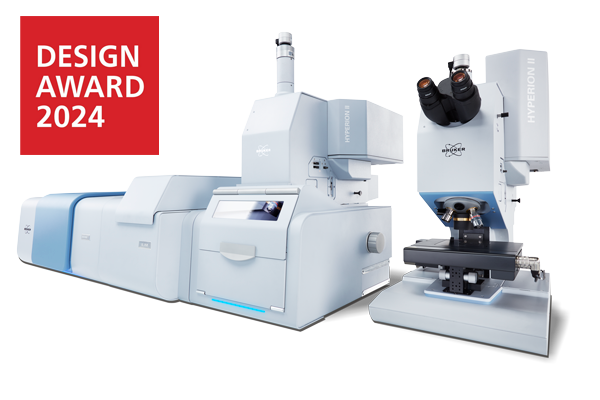
HYPERION II
Combinaison IRTF et QCL
Puissance analytique et innovation
La plateforme de recherche en Microscopie et Imagerie IR
Quoi de neuf avec HYPERION II:
Comment nous avons intégré l’imagerie laser infrarouge :
Notre réduction brevetée de la cohérence spatiale :
Microscopie IRTF amélioré par QCL | Imagerie laser infrarouge
L’HYPERION II est une innovation majeure en microscopie infrarouge. Il fournit une imagerie IR à la limite de la diffraction et établit la référence en microscopie ATR. Il combine pour la première fois la microscopie IRTF et la microscopie par imagerie laser infrarouge (ILIM) dans un seul appareil, offrant les trois modes de mesure: transmission, réflexion et ATR.
Caractéristiques de l’HYPERION II :
Sélection de différents détecteurs pour la microscopie IRTF:
Détecteur MCT refroidi LN2 large, moyen et bande étroite et MCT thermoélectrique (TE).
Détecteur FPA pour l’imagerie infrarouge (64 x 64 ou 128 x 128 pixels).
En option, ajout du module QCL pour l'imagerie infrarouge laser (ILIM, classe laser 1)
Sélection de différents objectifs: 3.5x / 15x / 36x / 74x IR, 20x ATR, 15x GIR, 4x / 40x VIS.
Extension de la gamme spectrale – du proche infrarouge (NIR) au lointain infrarouge (FIR)
Sélection des diaphgrames: couteaux manuels, roue de diaphgrame automatisée à couteaux. Diaphgrames métalliques pour le Proche IR
Sélection de différents accessoires et de table : accessoire d’imagerie macro IR, Table avec système de refroidissement/chauffage, compartiment d’échantillon, etc.
Outils pour l'amélioration du visuel/optique : éclairage darkfield, éclairage en fluorescence, polariseurs VIS, polariseurs IR, etc.
L'HYPERION II fournit :
Correspondance parfaite des images spectrales et visuelles. S’applique à n’importe quel mode de mesure (y compris l’imagerie ATR).
La diffraction a la limite en microscopie et imagerie IRTFavec une haute sensibilité à l’aide d’un détecteur FPA (Focal Plane Array).
En option, première combinaison de la technologie IRTF et QCL avec le module d’imagerie laser infrarouge (ILIM, classe laser 1).
Imagerie laser infrarouge dans tous les modes de mesure (ATR, Transmission, Réflectance).
Réduction de la cohérence brevetée pour des mesures d’imagerie laser sans artefacts sans perte de sensibilité ou de vitesse.
Vitesses d’imagerie élevées :
0,1 mm2 par seconde (FPA, spectre complet)
6,4 mm2 par seconde (ILIM, un nombre d’onde)
En option, le détecteur TE-MCT pour effectuer la microscopie IR avec une résolution spatiale et une sensibilité élevées sans azote liquide.
En option, la mesure d’émission et extensions de la gamme spectrale.
Applications avec le microscope HYPERION II :
Sciences de la vie et imagerie cellulaire
Produits pharmaceutiques
Études d’émissivité (p. ex. LED)
Analyse de défauts
Science médico-légale
Microplastiques
R&D industrielle
Polymères & Plastiques
Caractérisation de surface
Semi-conducteur
Le microscope IRTF de recherche pour l'innovation et la recherche de pointe
L'HYPERION II est le microscope qui incarne nos utilisateurs comme cela n'a jamais été le cas précédemment :
Flexible, précis, configurable, adaptable et toujours à la limite du possible.
Prenez le contrôle total
Il s’agit avant tout d’avoir un accès complet à un instrument. Accès à l'expérience, aux échantillons et aux paramètres. C'est la base de l'HYPERION II et son atout le plus précieux : offrir un contrôle total.
Qu'il s'agisse de mesures FT-IR en mode point unique, de cartographie ou d'imagerie avec différents détecteurs ou objectifs, des platines d'échantillonnage spéciales ou avec un objectif ATR ou Grazing Angle. À tout moment, vous pouvez influencer le résultat de vos résultats et les améliorer.
C'est la nette différence avec notre microscope IR LUMOS II. Là où le LUMOS II soulage l'utilisateur des détails expérimentaux fastidieux et automatise le processus de mesure, l'HYPERION II reste un outil précis qui ne fait que ce que l'utilisateur demande.

Une référence évolue
De nombreux utilisateurs connaissent l’HYPERION II et ses points forts grâce à son prédécesseur, l'HYPERION. Depuis près de 20 ans, il est une force d’innovation en microscopie et imagerie IR.
L’HYPERION II propose les mêmes fonctionnalités dont vous avez besoin dans votre routine et en recherche quotidienne: Détecteur MCT refroidi avce l'azote liquide et thermoélectriquement, détecteurs imagerie FPA, outils d’amélioration visuelle et infrarouge, et bien sûr une abondance d’accessoires dédiés.
En fin de compte, nous voulions établir une fois de plus la référence en matière de microscopie et d’imagerie IRTF et être à la hauteur de l'image de Bruker en tant que leader en innovation en introduisant une nouvelle technologie passionnante tout en conservant des méthodes établies et appréciées.

Augmentation de l'imagerie IRTF avec la technologie QCL
QCL et IRTF dans un seul instrument
Pour la première fois, les utilisateurs peuvent accéder à un microscope IR qui combine les technologies IRTF et QCL dans un seul instrument. Avec cela, nous ouvrons une toute nouvelle porte vers les sciences de la vie et la recherche sur les matériaux.
Collectez un spectre IRTF, sélectionnez les longueurs d’onde que vous souhaitez étudier à l’aide du QCL et créez des images chimiques époustouflantes en quelques secondes.
Avec cette toute nouvelle approche de l’imagerie laser IRTF et infrarouge, nous donnons enfin aux utilisateurs, aux chercheurs et aux scientifiques un outil pour développer de nouvelles applications, mais aussi pour améliorer les approches établies et éprouvées.
Un véritable microscope QCL aux performances exceptionnelles
L’HYPERION II propose un microscopie QCL sans compromis dans un microscope IRTF de pointe. En fait, nous avons spécifiquement développé et breveté une nouvelle technologie de réduction de cohérence pour permettre des performances d’imagerie laser IR inégalées - sans post-traitement numérique.
Pour illustrer : En spectroscopie IRTF classique, la cohérence spatiale ne joue aucun rôle. Dans les mesures en microscopie IR avec un QCL, cependant, des phénomènes de cohérence spatiale se produisent inévitablement. Ces franges et tavelures dans les images et les spectres IR sont généralement considérées comme gênantes pour l’imagerie chimique (voir ci-contre; DOI: 10.1002/jbio.201800015).
En effet, il n’est pas facile de séparer l’information chimique et physique de l’échantillon en décrivant la relation de phase des photons diffusés. L’HYPERION II résout ce problème de manière pragmatique et le résout par une conception matérielle intelligente et vous permet d’acquérir des données d’imagerie chimique sans artefacts.
Comparaison de la spectroscopie IRTF et QCL
Comparer les techniques impliquerait que les deux peuvent effectuer la même tâche aussi bien - une idée populaire qui est fausse. L’imagerie IRTF et par laser infrarouge présente des avantages distincts et seule une combinaison pratique des deux techniques permet d'obtenir les meilleurs résultats.
Nous savons que la plupart des scientifiques et des chercheurs ne veulent pas manquer l’universalité de l'IRTF et avoir des limites avec une seule technique de pointe. Heureusement, l’HYPERION II peut être considéré à la fois comme un microscope d’imagerie IRTF exceptionnel et un microscope QCL ambitieux.
Nous avons abordé cette dualité et là où la technologie QCL enregistre des données beaucoup plus rapidement avec un signal sur bruit identique, elle est encore limitée à une petite portion du domaine spectral du MIR. Encore une fois, nous restons fidèles au concept de l’HYPERION II. C’est vous qui choisissez. Vous avez le contrôle total.


Applications en microscopie IR (FPA, MCT, QCL)
Analyse biologique de tissus

Science des matériaux

Sciences médico-légales

Développement de médicaments

Géologie et minéralogie

Analyse des microplastiques

Matériaux énergétiques / Cristaux photoniques

Dans l'étude d'Arpin et al. (2013), les cristaux photoniques de tungstène auto-assemblés en 3D conservent leur structure jusqu'à environ 1 400 °C, tout en présentant des spectres de réflectance/émissivité IR adaptés qui montrent une émission thermique utile améliorée et une émission IR indésirable supprimée.
Rôle de l'HYPERION: Les chercheurs ont utilisé la réflectance IR pour déduire l'émissivité spectrale via la loi de Kirchhoff et ont appliqué l'analyse de réflectance pour établir un lien direct avec le contrôle de l'émissivité des cristaux photoniques en tungstène.
Nanophotonique / Bioanalyse

Amenabar et al. (2013) ont démontré la cartographie par nanospectroscopie infrarouge (nano-FT-IR) de complexes protéiques individuels (par exemple, la ferritine, les fibrilles d'insuline) avec une résolution latérale d'environ 30 nm, interprétant les spectres à large bande locaux dans la région amide I/II pour déduire la structure secondaire à l'échelle nanométrique.
Rôle de l'HYPERION: Les spectres IRTF ont été acquis comme spectres de référence et pour comparer les positions des pics et les formes des bandes avec les données nano-IRTF, soutenant ainsi les attributions des résonances de structure secondaire locale.
Plasmonique / Super Cristal

Mueller et al. ont synthétisé des « supercristaux » de nanoparticules d'or tridimensionnels auto-assemblés et ont démontré que les polaritons plasmoniques dans ces cristaux induisent une forte amplification du champ proche sur les bandes visibles et infrarouges moyennes, permettant à la fois la spectroscopie SERS et SEIRAS des molécules de ligand.
Rôle de l'HYPERION: Les spectres de réflectance et de transmittance IR sur des films de supercristaux plasmoniques ont été collectés, démontrant le couplage entre les modes plasmoniques et les vibrations moléculaires et quantifiant l'amélioration SEIRAS.
Dépistage sur puce / Biomédical

Dans Benz et al. (2020), une lame d'oxyde d'indium-étain (ITO) à motifs à base de dendrimère a été utilisée pour générer plus de 50 000 nanorécipients par plaque, permettant la synthèse chimique sur puce et la spectrométrie de masse MALDI-TOF ultra-sensible, la surveillance de la réaction UV-Vis (RM) et la spectroscopie IR sur puce pour la RM et la caractérisation dans la découverte de médicaments à haut débit.
Rôle de l'HYPERION: Les spectres IR des points du réseau de gouttelettes ont fourni des signatures d'absorption vibrationnelle moléculaire pour compléter les données MS et UV-Vis, soutenant ainsi la caractérisation à haut débit de la bibliothèque synthétisée.
Plasmonique / Métamatériaux

Paggi et al. (2023) démontrent un résonateur métasurface surcouplé qui améliore l'absorption vibrationnelle moléculaire dans la gamme infrarouge moyenne (5–10 μm) en couplant des modes plasmoniques faibles aux vibrations moléculaires, atteignant des variations de réflectivité jusqu'à 1 % par nanomètre d'épaisseur de couche moléculaire.
Rôle de l'HYPERION: Les spectres IR dans l'infrarouge moyen ont permis d'évaluer l'influence du résonateur métasurface sur les modes de vibration, d'éclairer le mécanisme SEIRA à large bande et de soutenir la conception de plateformes de détection.
Polariscopie / Nanomatériaux

Honda et al. (2019) ont démontré la polariscopie infrarouge (IR) par imagerie hyperspectrale avec un détecteur à réseau de plans focaux (FPA) dans la région spectrale IR, sous illumination par des sources de lumière thermique et synchrotron. Ils montrent que le temps nécessaire à la polariscopie aux longueurs d'onde IR peut être considérablement réduit grâce à l'imagerie FPA-FTIR.
Rôle de l'HYPERION: Les expériences FPA-IRTF hors ligne et l'imagerie hyperspectrale basée sur le synchrotron ont toutes deux été réalisées sur l'Hyperion, permettant l'analyse des caractéristiques sub-longueur d'onde ciblées par polariscopie.
Matériaux énergétiques / Métamatériaux

Dyachenko et al. présentent la conception et la fabrication d'un métamatériau multicouche réfractaire composé de tungstène et de dioxyde d'hafnium, présentant une transition topologique optique entre une dispersion epsilon-proche-de-zéro et une dispersion hyperbolique. Cette transition permet de contrôler les propriétés d'émission thermique du matériau à haute température, autorisant ainsi un réglage sélectif de l'émissivité.
Rôle de l'HYPERION: Des spectres de réflectance ont été collectés dans la région NIR afin de déterminer la position spectrale et l'intensité des résonances optiques dans la structure multicouche.
Optique / Imagerie IR

Stavitski et al. (2013) ont atteint une résolution spatiale limitée par la diffraction en couplant des lignes de faisceau infrarouges (IR) synchrotron à des détecteurs à réseau de plans focaux (FPA), permettant ainsi l'imagerie plein champ avec une sensibilité chimique élevée et des temps d'acquisition rapides. Grâce à cette approche, ils ont cartographié avec succès la composition chimique d'échantillons hétérogènes avec une résolution spatiale d'environ 5 µm.
Rôle de l'HYPERION: Doté d'un détecteur FPA, l'HYPERION a servi de plateforme d'imagerie couplée au synchrotron utilisé pour l'analyse chimique et compositionnelle.
Catalyse / Matériaux énergétiques

Cao et al. rapportent l'utilisation du rayonnement synchrotron operando FT-IR (SR-FT-IR) sous des potentiels de travail pour détecter une bande d'absorption vibrationnelle réversible à ~764 cm⁻¹ d'un catalyseur Ru₁–N₄ atomiquement dispersé, qu'ils attribuent à l'adsorption/désorption d'un intermédiaire d'oxygène (O*), révélant ainsi l'implication dynamique des espèces O sur le site Ru pendant la catalyse.
Rôle de l'HYPERION: Un dispositif SR-IRTF operando a été utilisé pour interroger spatialement et spectroscopiquement le catalyseur sous polarisation, capturant la signature vibrationnelle de O* sur le site Ru et confirmant le lien direct entre l'adsorption intermédiaire et la fonction catalytique.
Catalyse / Matériaux énergétiques

Dans l'étude de Su et al., les auteurs conçoivent des agrégats de CuO supportés sur du carbone dopé à l'azote et démontrent que, sous l'effet d'un potentiel appliqué, ces agrégats se restructurent dynamiquement en espèces Cu₂–CuN₃ présentant une asymétrie de charge, ce qui est corrélé à une efficacité faradique élevée pour la production de composés C₂⁺ (notamment l'éthanol). Ils combinent les techniques XAS operando, XPS quasi-in situ et FT-IR operando (avec calculs DFT) pour suivre en temps réel les modifications structurales, électroniques et d'adsorbat.
Rôle de l'HYPERION: Dans le cadre de leur dispositif IRTF operando, le faisceau infrarouge synchrotron a été focalisé dans la cellule de réaction afin de recueillir les spectres de réflectance sous différents potentiels. Ces mesures permettent de suivre les caractéristiques vibrationnelles moléculaires et les signatures d'asymétrie de charge liées aux agrégats Cu₂–CuN₃ et ainsi de confirmer les corrélations structure-activité en conditions opératoires.
Accessoires pour l'échantillonnage et Objectifs
Plaque de validation

Porte-échantillon MicroVice

Accessoire d’imagerie macro ATR

Porte-filtre

Support chauffant de de refroidissement

Objectif d’imagerie 3,5x

Objectifs à transmission standard

Objectif ATR

Objectif à angle rasant

Cellule de compression diamant

OPUS Release 9.0 Highlights for HYPERION II | Q1 2025
New Feature: The Autonomous Composition Identifier (A.I.D.) simplifies search and identification.
The Autonomous Composition Identifier (A.I.D.) is an AI-driven software tool for identifying the chemical composition of samples based on IR or Raman spectral data. It incorporates the previous O/SEARCH features and adds a multi-step algorithm to find the best match from reference spectral databases, whether dealing with pure substances or complex mixtures. A.I.D. intelligently evaluates and recommends optimal matches with a balance of reliability and sensitivity—automatically generating diagnostics and visual reports, including residual spectra and hit quality graphs. It delivers high-performance results in seconds and supports in-depth, interactive analysis of alternative identifications.

New Feature: Co-local Measurement – Unified Analysis Across IR and Raman
The Co-local Measurement function, part of the OPUS/OBJECT package, enables seamless analysis of the same region of interest on microscopic samples using both IR and Raman microscopy. It allows the transfer of overview images and measurement areas between LUMOS II, HYPERION II, and SENTERRA II instruments. This ensures that IR and Raman data are collected from precisely the same spot, enabling complementary chemical insights. Co-local Measurement bridges the gap between two techniques, improving data correlation and boosting confidence in results for heterogeneous or complex samples.

Updated Feature: OPUS/OBJECT now combines "Find Particle" and "Cluster ID"
The Particle Identification function (part of the OPUS/OBJECT package) combines the power of “Find Particles” and “Cluster ID” into one streamlined workflow. It detects particles within IR and Raman chemical images, determines their dimensions, and identifies their chemical makeup—all in one go. This integrated approach simplifies analysis and accelerates decision-making for complex samples.

Parution de OPUS 8.7 | HYPERION II | Q3 2021
Nouvelles caractéristiques: Génération d'images chimiques hautes performances grâce à la nouvelle fonction de clustering adaptatif K-means
Cette nouvelle fonction est la nouvelle étape de développement logique de notre fonction d'analyse de cluster bien connue. La fonction de clustering adaptatif K-means est basée sur un nouvel algorithme, qui permet une détermination non supervisée et autonome de la variance spectrale dans vos résultats d'imagerie ou de cartographie..
- La prévision ou la recherche fastidieuse de la quantité de classes chimiques incluses n'est plus nécessaire car l'algorithme peut prédire automatiquement toutes les classes chimiques.
- Cette fonction majeure est importante pour tous les types d'imagerie chimique et d'analyse de distribution d'échantillons inconnus ou de petites structures dans des ensembles de données importants.
- Rendre l'analyse et l'évaluation très simple et économiser votre temps.
Nouvelle fonction: “Fonction « Cluster ID » pour l'identification des classes dans les données spectrales 3D
Notre nouvelle fonction Cluster ID permet l'identification de clusters dans des données imagerie et de cartographie à l'aide des fonctions OPUS : recherche de spectre dans les bibliothèques, comparaison rapide ou test d'identité.
- Détermination facile de l'identité chimique des composants d'échantillons classifiés pour les particules, les multi-couches, les composants de comprimés pharmaceutiques et d'autres matériaux non homogènes.
- Des rapports statistiques fiables et complets sur la quantité, la taille et bien sûr l'identité de toutes les structures analysées sont fournis et conduisent l'analyse des particules à un nouveau niveau autonome.
Evolution de fonction: "La fonction " Find particles" contient une nouvelle méthode de détection des particules
La fonction "Find Particle" peut maintenant être appliqué simultanément à l'image visuelle et IR. Avec cette mise à jour de la fonctionnalité, vous pouvez effectuer une détection de particules basée sur des images chimiques qui ont été mesurées par le LUMOS II.
- Alors que la reconnaissance des particules pour les structures à faible contraste et les particules/fibres blanc cassé/transparentes sur les filtres 'blanc cassé' peut être fastidieuse, une détermination des particules post-analyse basée sur l'image IR chimique vous permet de déterminer la quantité et la taille des particules à partir de votre imagerie ou cartographie.


Demande d'informations complémentaires
En savoir plus sur nos microscopes et solutions IR-TF en téléchargeant de la documentation associé.


