
Linking Cardiac Nanostructure and Molecular Organization to Cardiac Function
Using EM and SMLM for novel insights into the heart
Attendees will learn how to relate the nanoscale structural analysis of cells and tissues to their function with special focus on:
- Using indirect Correlative Light and Electron Microscopy (iCLEM) to correlate EM and SMLM data.
- How our guest speakers use iCLEM to study the nano-structural underpinnings of electrical signal propagation in the heart.
- Learning how to interpret quantitative data and incorporate it into computational models.
- How research can provide a basis for the development of novel classes of anti-arrhythmic drugs.
Discover the Latest Advances in Structural Analysis at the Nano Scale
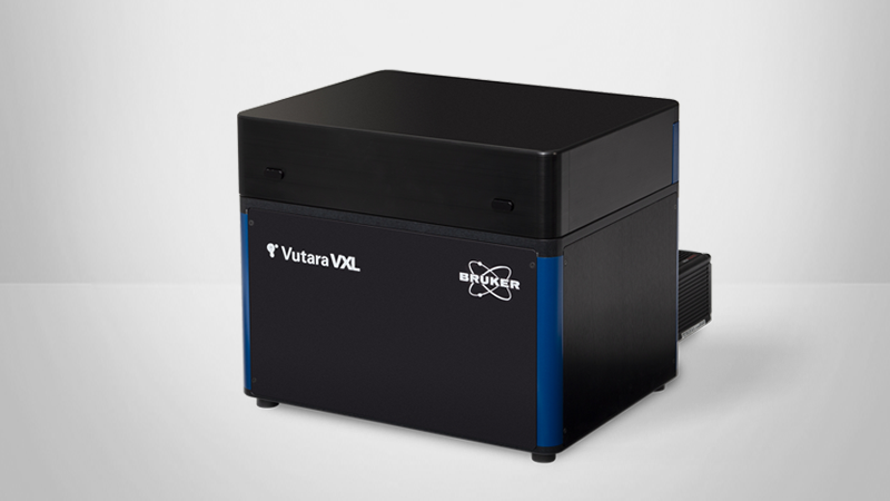
Correlative light and electron microscopy (CLEM) is emerging as a powerful tool for nanoscale structural studies in both the biological and physical sciences. However, the challenges associated with obtaining precisely registered images of nanoscale neighborhoods within a sample on multiple microscopes severely limit the adoptability and throughput of CLEM. Additionally, the low throughput and lack of robust image analysis led to CLEM being largely used as a qualitative approach with limited ability to assess structural heterogeneity or identify subtle changes associated with the early stages of disease. By leveraging the high throughput of the Vutara single molecule localization microscope and taking advantage of structural fiducials/landmarks, identifiable via both light and electron microscopy, we have developed indirect CLEM (iCLEM) as a low-cost, high throughput option with extensive quantitative capabilities.
This approach has enabled us to undertake a systematic investigation of the structural underpinnings of electrical signal propagation in the heart, a critical process that coordinates the contraction of ~12 billion muscle cells during each heartbeat. Motivation for this work derives from the fact that disruption of this process leads to life-threatening disruptions in the heart’s rhythm (arrhythmias). Therefore, we used iCLEM to study the intercalated disk (ID), a specialized structure that affords electrical and mechanical coupling between muscle cells in the heart. Although the ID is widely recognized as a complex, heterogeneous structure, the lack of quantitative structural data has forced computational models to omit it or simplify it to a homogenous cylinder. Using iCLEM, we have obtained the first-ever quantitative picture of the ID, enabling us to construct realistic computational models. Transmission electron microscopy (TEM) enabled us to quantify ID ultrastructure from micro- through nano-scales and thereby, construct populations of finite element models of IDs, capturing both intra- and inter-individual variability. We then populated these meshes with electrogenic proteins based on STORM-based Relative Localization Analysis (STORM-RLA) of single-molecule localization data. By incorporating these data into computational models of electrical signal propagation, we are uncovering previously unappreciated structure-function relationships that determine the regularity of the heart’s rhythm. These predictions, along with functional imaging studies of electrical signal spread in the heart, are providing the basis for the development of novel classes of anti-arrhythmic drugs.
Find out more about our other solutions for Super-Resolution Microscopy:
Guest Speakers
Rengasayee (Sai) Veeraraghavan, Ph.D.
Rengasayee (Sai) Veeraraghavan, PhD, is an Assistant Professor in the Department of Biomedical Engineering at the Ohio State University (OSU). His lab (Nanocardiology lab, https://nanocardiology.osu.edu/, https://twitter.com/nanocardiology) uses light and electron microscopy techniques and computational image analysis approaches to uncover how nanoscale structure influences the heart’s rhythm in health and disease with the goal of developing novel therapies against life-threatening rhythm disturbances (arrhythmia). His research is currently supported by grants from the National Institutes of Health and the American Heart Association.
Heather Struckman, BS, MS
Heather Struckman, BS, MS, is a graduate student in the Nanocardiology lab who is applying confocal microscopy, STORM, STED, transmission electron microscopy, and optical mapping (high-speed imaging of electrical signals from live hearts) to investigate the structural basis of cell-to-cell electrical communication in the heart. She is a National Science Foundation Fellow entering the fourth year of her doctoral training in Biomedical Engineering at the Ohio State University. So far, she has published 6 peer-reviewed articles and presented over 20 abstracts at national/international meetings (including 8 oral presentations), and won a $20,000 Strategic Initiative Grant from the Microscopy Society of America for her outreach efforts.

