
Single-Molecule Localization Microscopy: Beyond the Coverslip
Discover the impressive insights that SMLM can bring to your lab
During this webinar, attendees will gain first-hand insights into the working principles and applications of single-molecule localization microscopy (SMLM) with Bruker’s Vutara super-resolution microscope. The Vutara uses bi-plane detection and widefield illumination to give users the flexibility to image various sample types including:
- Cell cultures
- Tissue slices
- Whole organisms
- Hydrogels
- Purified samples (proteins, DNA/RNA, or viruses)
Webinar Summary
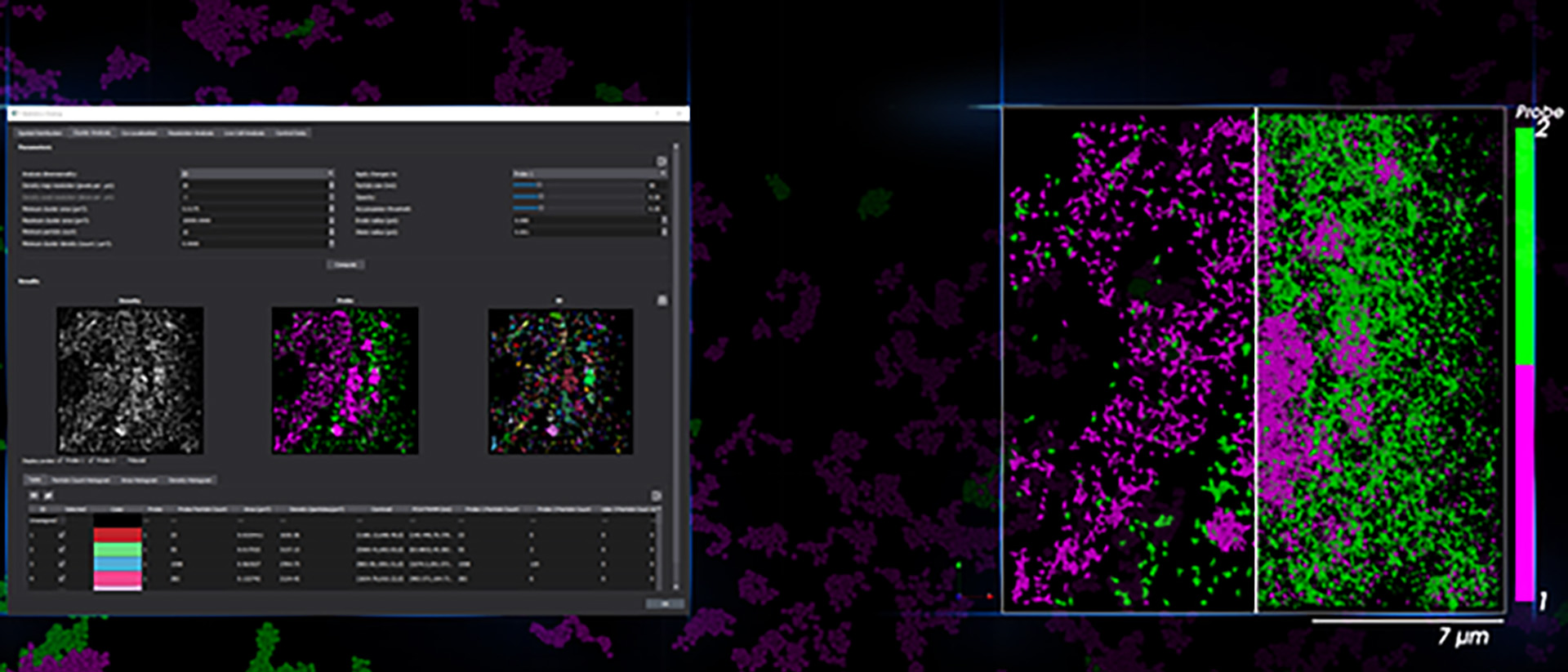
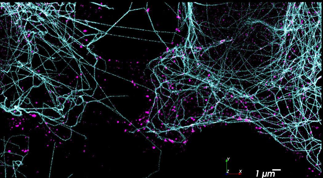
Single-molecule localization microscopy (SMLM) provides the ability to image biological processes well below the diffraction limit of light. Commercial and homemade systems for SMLM typically utilize TIRF based illumination which limits the depth of imaging to a few microns from the cover slip. This restricts the range of detection to only part of cultured adherent cells and makes imaging of cell colonies, tissues, or small organisms impractical.
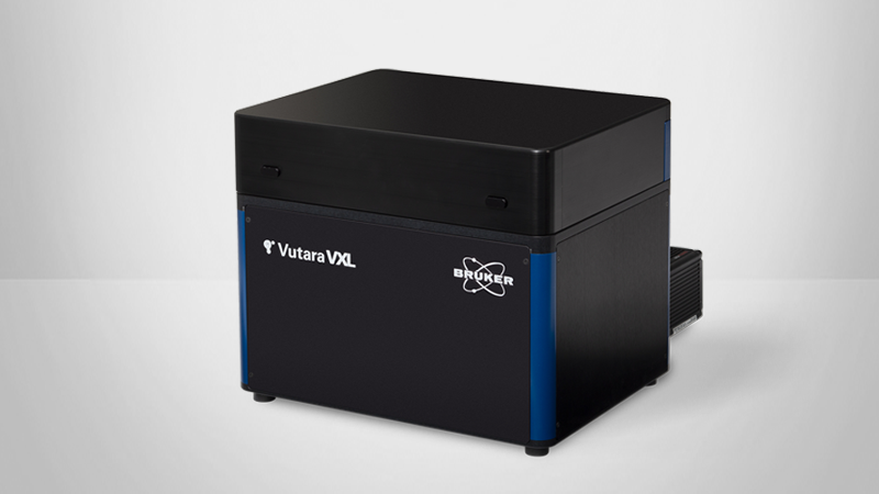
The Vutara SMLM system is not constrained by this limitation and is capable of imaging throughout the entire axial dimension of cultured adherent cells, tissues, and small organisms by utilizing biplane detection along with widefield illumination. This webinar will describe the technical details of Vutara, as well as provide application examples demonstrating the use of the Vutara for various sample types and SMLM techniques such as STORM and DNA PAINT.
Featured Products and Technology

Speaker

Dr. Lauren Gagnon
Vutara Applications Scientist, Bruker
Dr. Lauren Gagnon graduated from Emmanuel College with a degree in Chemistry before completing her PhD in Chemistry at the University of Washington. Her thesis research focused on the development of novel approaches to single molecule localization microscopy using DNA barcoded antibodies.