

Luxendo Light-Sheet Technology
Selecting a Light-Sheet System
Can't find the answer to your question? Feel free to contact us. We will be happy to connect you with a light-sheet microscopy expert who can assist you with any questions you have.
General
What is light-sheet fluorescence microscopy?
Light-sheet fluorescence microscopy (LSFM), or single-plane illumination microscopy (SPIM), is a microscopy technique used for a variety of samples across scales:
- Live, fixed, or cleared samples
- Large samples and whole-body specimens
- Fast dynamic biological processes
- Live specimens over long periods
How does a light-sheet microscope work?
Light-sheet fluorescence microscopes (LSFM) decouple the fluorescence excitation and the detection beam path geometrically, and not spectrally as in widefield or confocal fluorescence microscopes. LSFM is a microscopy method in which a sample is illuminated with a thin sheet of light in the image plance of the detection objective lens, therefore avoiding the generation of out-of-focus fluorescence signal. This is a major difference to confocal microscopy where effectively the whole sample volume is illuminated, so that out-of-focus fluorescence signal is generated but then rejected by the detection pinhole. The optical setup and use of a thin sheet of laser light enables inherent optical sectioning.
The emitted fluorescence signal of the entire illuminated 2D plane is simultaneously detected by a scientific camera, typically scientific CMOS (sCMOS) cameras, rather than pinhole-based selection and photomultiplier tube (PMT)-based detection of light from a single pixel at a time. Therefore, LSFM allows for fast imaging with high temporal and 3D-spatial resolution. It also shows significantly reduced phototoxicity and photobleaching, high imaging speed, and low background.

What are the advantages of using a light-sheet microscope compared to a confocal microscope?
Light-sheet fluorescence microscopy (LSFM) is significantly faster and less phototoxic than conventional laser point-scanning techniques.
Light-sheet imaging is often used for one or several of these reasons:
- Reduced phototoxicity and photobleaching: Light-sheet imaging minimises light exposure to the specimen by selectively illuminating a single plane. This makes it suitable for imaging live samples over extended periods.
- High speed: Light-sheet microscopy can acquire 3D image stacks rapidly, making it ideal for capturing dynamic processes in live samples.
- Low background: Since out-of-focus light is not detected, light-sheet microscopy produces images with high contrast and minimal background.
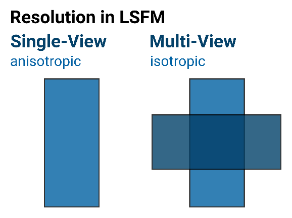
How can light-sheet microscopy improve 3D image resolution of biological tissues?
In fluorescence microscopy, the axial resolution is often lower than the lateral resolution due to light interaction with matter and optical geometries. This results in anisotropc point spread functions (PSF), which means that the resolution is anisotropic.
Using a multi-view light-sheet microscope enables samples to be imaged from different angles, and applying subsequent image processing steps, i.e., image registration, fusion, and deconvolution allows to create isotropic data, which have equal resolution in x, y, and z. The LuxProcessor image processing software, part of the LuxBundle software package, has been developed for exactly this type of image processing of multi-view light-sheet data with a focus on ease of use.
About Luxendo Light-Sheet Microscope Technology
Can I image live and cleared samples using one Luxendo microscope?
Yes, the Luxendo MuVi SPIM can support cleared, fixed, and live sample imaging by a simple exchange of the central optical unit, called the octagon.
This exchange takes only a few minutes and gives you the flexibility to image live and cleared samples on the same microscope.
Can the Photomanipulation Module be added to all Luxendo light-sheet microscopes?
The photomanipulation module can be added to the Luxendo light-sheet microscopes designed for live imaging, namely:
Luxendo light-sheet microscope systems are designed to provide high modularity and can be easily adapted to suit the requirements of your experiment. Contact us to discuss your specific needs with a light-sheet microscopy expert and to receive recommendations and guidance tailored to your experiments.
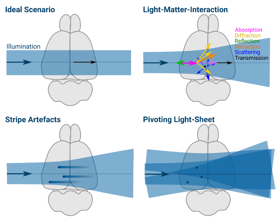
How does Luxendo light-sheet technology mitigate striping artifacts?
Bruker's Luxendo light-sheet microscopes achieve destriping by pivoting the illumination light-sheet around the center of its beam waist. High-end precision is achieved using a tube lens and an ultrafast scanning mirror, which deflects the laser beam, allowing the light beam to illuminate all points of the sample with a broad range of angles as it oscillates. As the oscillation is much faster than the camera exposure time, a homogenous illumination profile is generated, thus minimising potential striping artefacts without compromising acquisition speed.
How long can a Luxendo light-sheet microscope with environmental control image live samples?
The Luxendo environmental control module ensures precise management of environmental parameters essential for both long-term and live imaging. Luxendo light-sheet microscopes have been used for multi-day continuous acquisitions on live samples lasting up to 7 days.
Temperature Regulation: Utilizing a Peltier-based water cooling and heating unit, temperatures can be finely tuned within the range of 20°C to 39°C. This capability establishes ideal incubation conditions suitable for a diverse array of biological specimens, spanning from marine organisms to mouse embryos and organoids.
Gas Concentration Control: Gas concentrations for various components are customizable, allowing for tailored settings:
- CO2 level adjustable from 0% to 15%.
- Oxygen (O2) concentration adjustable from 1% to 21%.
- Humidity (H2O) level adjustable from 20% to 99%.
How are such vast data sets managed?
Luxendo light-sheet microscopes are compatible with Acquifer HIVE, our comprehensive data processing and storage solution. HIVE data management systems feature a fast backbone for very high data collection speed and multicore, multi-GPU processing, as well as scalable plug-and-play storage modules up to the petabyte (PB) range.
Moreover, Acquifer HIVE is designed to manage the vast amounts of biological imaging data as well as data from high-content screening, sequencing, multi-omics/spatial biology, and other data structures.
What image processing tools are available with Luxendo light-sheet microscopes?
Light-sheet fluorescence microscopy generates large image data, often multiple terabytes, that require efficient processing software to give users fast results. Therefore, efficient software is constantly evolving to be able to handle and process light-sheet data. Bruker's LuxBundle software for Luxendo light-sheet microscopes integrates:
- LuxControl for flexible microscope control
- LuxViewer for image viewing
- LuxProcessor for powerful post-processing
Luxendo's software provides a 3D-data viewer, tile stitching, multi-view fusion, and deconvolution as image post-processing tools to deliver whole 3D volume datasets that are ready to use for your image analysis pipeline.
Do I need a specialized room for my Luxendo light-sheet microscope?
No, Luxendo light-sheet microscope microscopes are designed to be compact, robust, and vibration-free. This allows you to operate Luxendo light-sheet microscopes on any sturdy table (e.g., close to the lab bench) and in a daylight room. Additionally, Luxendo light-sheet microscopes have a fully enclosed optical beam path. Thus, all Luxendo systems are certified as Laser Safety Class 1 systems, i.e., the same safety class as a laser pointer.
See this question answered in more detail during the audience Q&A session at the end of the light-sheet segment of our on-demand webinar "Bruker Microscopes Fill the Gap Between Traditional Fluorescence and Electron Microscopy" to see this question discussed in greater detail.
Choosing A Light-Sheet Microscope
What are some general considerations?
Choosing a microscope depends on several factors, such as sample type, sample size, resolution requirements, field of view (FOV), mounting needs, and imaging speed. The following segments provide some general guidelines for choosing the right Luxendo light-sheet microscope.
Contact us to discuss your specific measurement requirements with a light-sheet microscopy expert. We will be happy to provide instrument and configuration recommendations tailored to your scientific needs.
System Comparison
* COMPATIBLE MODULE
| The perfect allrounder - multi-view imaging for diverse samples (live and cleared option) | Multi-sample and dual view flexibility for highly sensitive imaging, ideal for organoids and 3D cell cultures | In vivo multi-sample and multi-condition imaging, ideal for drug treatment comparison | Perfection for cleared samples prepared by any type of clearing protocol | High-end imaging with various beam patterns from single cells to 3D cell cultures | |
|---|---|---|---|---|---|
| Geometry | Horizontal with multiview | Horizontal with dual view | Inverted with dual illumination | Inverted dual illumination with moving optics | Inverted geometry at 360° |
| Beam Type | Gaussian | Gaussian and Bessel | Gaussian | Gaussian | Advanced Illumination Module (AIM) |
| Application Examples | - Long-term imaging of Drosophila development - Photomanipulation studies in zebrafish embryos - Cleared and stained samples up to mouse brain size | - High-resolution full 3D imaging of multiple organoids over time - Long-term imaging of highly light-sensitive samples - Comparing the pharmacological impact in zebrafish wound healing after PM | - Time-lapse imaging of multiple pancreatic spheres - Studying pharmacological impact on zebrafish development - Comparing genetically altered organoids | - Pharmacokinetics in mouse brain - Multi-stained rat brain - Whole mouse imaging | - Fast imaging of photo-sensitive 2D cell culture - Lattice imaging for enhanced resolution in 3D cell cultures - FCS and FLIM in cell culture |
| User Level | Beginner friendly | Beginner friendly | Beginner friendly | Intermediate | Advanced |
| # Lenses | 4 lenses (2 IO, 2 DO) | 3 lenses (1 IO, 2 DO) | 3 lenses (2 IO, 1 DO) | 3 lenses (2 IO, 1 DO) | 2 lenses (1 IO, 1 DO) |
| Multi-View | ✓ | ✓ | |||
| Live/Fixed Samples | ✓ | ✓ | ✓ | ✓ | |
| Cleared Samples | ✓ | ✓ | |||
| Expanded Samples | ✓ | ✓ | ✓ | ✓ | ✓ |
| Best Embedding | Capillary/FEP tube w/ agarose 3D stage | Custom-solution and FEP foil | TruLive3D dishes and FEP foil | Quartz-crystal cuvette | FEP foil; glass slides |
| Photomanipulation* | ✓ | ✓ | ✓ | ✓ | |
| Environmental Control* | ✓ | ✓ | ✓ | ✓ | |
| Destriping/Uniform Illumination* | ✓ | ✓ | ✓ | ✓ | ✓ (w/ Advanced Illumination Module) |
| Benchtop Design | ✓ | ✓ | ✓ | ✓ | ✓ |
What is the best light-sheet microscope for live samples?
Luxendo light-sheet microscopes are ideal for non-invasive long-term imaging of live samples across scales, ranging from subcellular structures to cells, tissues, organoids, and embryos. Light-sheet imaging is often used for one or several of these reasons:
- Reduced phototoxicity and photobleaching: Light-sheet imaging minimises light exposure to the specimen by selectively illuminating a single plane. This makes it suitable for imaging live samples over extended periods.
- High speed: Light-sheet microscopy can acquire 3D image stacks rapidly, making it ideal for capturing dynamic processes in live samples.
- Low background: Since out-of-focus light is not detected, light-sheet microscopy produces images with high contrast and minimal background.
The multi-view MuVi SPIM is one of the fastest multi-angle view systems on the market. Due to its modular concept, MuVi SPIM facilitates both, live-sample (LS) and cleared-sample (CS) imaging by an acquisition unit exchange.
The TruLive3D Imager is optimized for fast 3D multi-sample volume imaging of delicate live specimens in their native 3D environment. It is particularly well-suited for time-lapse and high-throughput imaging of 3D spheroids, organoids, 3D cell cultures, and small embryos.
The InVi SPIM Lattice Pro provides state-of-the-art light sheet technology for high-resolution imaging of live and fixed samples. It uses spatial light modulator technology (SLM) to generate a wide variety of illumination beam patterns, allowing for optimal illumination for each sample and application.
What is the best light-sheet microscope for cleared samples?
The LCS SPIM is a compact light-sheet microscope specifically developed for large optically cleared samples. Importantly, the LCS SPIM is compatible with all currently available clearing solutions. The LCS SPIM allows cleared sample imaging rom organs to whole organisms, facilitating the visualization of complete neuronal networks and vascular trees at cellular resolution.
The multi-view MuVi SPIM is one of the fastest multi-angle view systems on the market. Due to its modular concept, MuVi SPIM facilitates both, live-sample (LS) and cleared-sample (CS) imaging by an acquisition unit exchange.
What is the best light-sheet microscope for both live and cleared samples?
The Bruker Luxendo multi-view MuVi SPIM is not only one of the fastest multi-angle view systems on the market, but also incorporates years of Luxendo light-sheet experience and innovation. Due to its modular concept, MuVi SPIM facilitates both, live-sample (LS) and cleared-sample (CS) imaging by an acquisition unit exchange.
The functional flexibility of the MuVi SPIM instruments is made possible by Luxendo’s highly adaptable modular concept. Users can quickly and easily exchange the central unit called Octagon, which consistis of the illumination objective, detection objective, and the sample mounting chamber.
What is the best light-sheet microscope for large samples/whole-body specimens?
The MuVi SPIM is optimal for studying large live samples (e.g., drosophila or zebrafish embryos) or large cleared samples up to entire mouse brains, all of which can be resolved at subcellular resolution.
The LCS SPIM is a dedicated microscope for cleared sample imaging and optimized for very large specimens up to entire adult cleared mice. This enables visualization of structures like complete neuronal or vascular networks.
Ask A Question
Get direct answers from the Bruker light-sheet microscopy team.