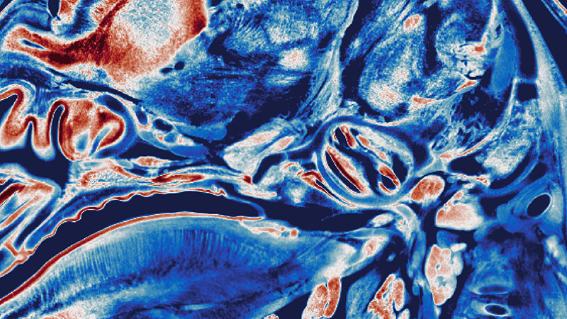
Cleared Sample Imaging
Cleared-Sample Imaging Solutions
Bruker Luxendo light-sheet microscopes are optimized for 3D imaging of intricate tissue structures and deliver best-in-class 3D imaging of cleared samples. The variety of Bruker Luxendo Selective-Plane Illumination Microscopes (SPIMs) allows researchers to choose the best microscope for their sample, depending on mounting, sample size, and optics. These characteristics are particularly important for complex studies such as brain connectomics or tumor metastasis.
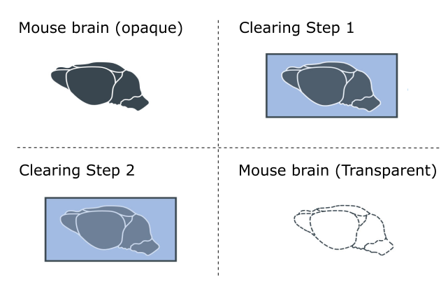
Tissue clearing in combination with light-sheet microscopy has become the go-to technique to visualize large, and by default, opaque tissues. Thanks to technological advancements, large structures can be imaged within minutes, providing new insights in the fields of neuroscience and developmental biology. Furthermore, as tissue clearing is a rapidly evolving field, Bruker Luxendo SPIMs are compatible with all clearing solutions available.


Cleared Sample Imaging Gallery
For more information about cleared sample imaging with light sheet fluorescence microscopy, visit:

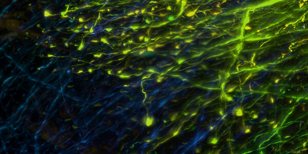
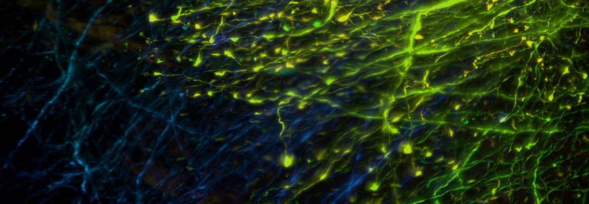
Neurons of a transgenic mouse expressing fluorescent protein YFP. Brain was cleared with Clarity.
Courtesy of:
Dr. Zhang Dan, Tsinghuan University, China
Cleared Mouse Lymph Node
MuVi SPIM
Cleared mouse lymph node. High endothelial venules (642 nm, red) and autofluorescence (488 nm, green) to visualize surrounding tissue. Imaged on the MuVi SPIM.
Courtesy of:
Jens Stein, University of Bern, Bern, Switzerland
Cleared Mouse Lymph Node
QuVi SPIM
BABB-cleared mouse lymph node. High endothelial venules (red) and autofluorescence (green) to visualize surrounding tissue.
Courtesy of:
Jens Stein, University of Bern, Bern, Switzerland

Light-sheet images of a lung taken with the LCS SPIM
Courtesy of:
Ayelen Melina, Santamans Recchini, and Gaudalupe Sabio Buzo, Stress kinases in Diabetes, Cancer and Cardiovascular Disease Laboratory and Unit of Microscopy and Dynamic Imaging, Centro Nacional de Investigaciones Cardiovasculares Carlos III (CNIC), Madrid.

Cleared Mouse Head
Labeled with anti-tuj1 (green) to mark developing nerves and with anti-desmin (red) to mark differentiating muscles. Tiled image (6 × 5).
Imaged on the MuVi SPIM.
Courtesy of:
Glenda Comai, Institut Pasteur, Paris, France

Developing Nerves in a Whole Mouse Embryo
Developing nerves in a whole mouse embryo.
Sample cleared with DBE. Tiled image (3 x 4) acquisition. Scalebars: 1mm.
Imaged on the LCS SPIM.
Sample courtesy of:
James Muller, MSKCC, New York, USA

Neuronal Network
Neuronal network (stained with GFP) of a CUBIC-cleared mouse brain
Color-coded depth representation: maximal depth displayed 1.23 mm.
Imaged on the MuVi SPIM.
Courtesy of:
Montserrat Coll Lladó, European Molecular Biology Laboratory (EMBL), Barcelona, Spain