MuVi SPIM

MuVi SPIM
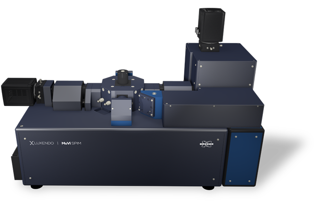
Bruker's Multi-View Selective-Plane Illumination Microscope (SPIM) is not only the fastest light-sheet system on the market, but also incorporates years of Luxendo imaging experience and innovation. Due to its modular concept, MuVi SPIM facilitates both live-sample (LS) and cleared-sample (CS) imaging by an acquisition unit exchange.
MuVi SPIM allows for high-speed volumetric acquisition of dynamic processes in live specimens, as well as cleared sample imaging with compatibility with any clearing method.
Best-in-Class Multiview Imaging Capabilities
Convenient Multiview Imaging
MuVi SPIM features a unique 4-axis concept and is available in various configurations to enable:
- Simultaneous illumination and detection from two opposing directions without rotation
- Unparalleled acquisition speed
- Free rotation axis for optimal sample positioning and 360° view
- Long-term stability for imaging time series over days
Isotropic Resolution with 360° Illumination and Detection
The MuVi SPIM is equipped with a motorized sample holder which enables sample rotation by 360°. Although sample rotation is not required to achieve in-toto imaging, a single 90° rotation of the sample can provide isotropic resolution and improve image quality. This is at least four times faster compared to conventional SPIM.

Next-Generation Technology
Built on Bruker's history of innovation, our next-generation MuVi SPIM incorporates several new features to enhance image quality, shorten acquisition speed, and allow high-resolution and artifact-free imaging of a broader range of samples.
New Advanced Features
Larger Field-of-View (FOV) and Superior Axial Resolution
When imaging, obstacles like pigments or cell nuclei can interfere with light, leading to images with shadows and striping artifacts. The next-generation SPIM employs dual de-striping via pivot scanning which generates a homogenous illumination profile without compromising acquisition speed.
Improved Signal-to-Noise
When imaging thicker samples or opaque specimens, light scattering can lead to reduced contrast and image blur. MuVi SPIM has implemented an elegant approach to enhance image contrast by offering:
- Scanned light-sheet
- Adjustable light-sheet thickness
- Robust aberration tolerance (even in complex samples)
- Line illumination for improved background suppression
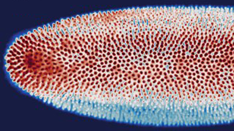
Dual-Destriping: Impressive Elimination of Striping Artifacts
When imaging, obstacles like pigments or cell nuclei can interfere with light, leading to images with shadows and striping artifacts. The next-generation SPIM employs dual de-striping via pivot scanning which generates a homogenous illumination profile without compromising acquisition speed.

TAG-Lens: Light-Sheet Length Control with Fast Axial Scanning
Thanks to state-of-the-art technologies, ultra-fast varifocal lenses can be used to fine-tune the system’s optics. Luxendo SPIMs use devices that are driven by an acoustic wave, called tunable acoustic gradient lenses (or TAG lenses), for adaptive optics. These TAG lenses allow better light-sheet control and uniformity while reducing axial stretching.


Transition between live sample and cleared sample system configurations in just minutes.
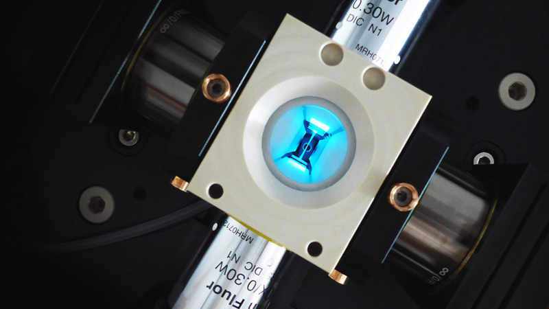
The sample chamber and all objective lenses are mounted on the octagon, the optical core unit that holds all the objective lenses.
Different octagons can support different magnifications and objective NAs and are compatible with objectives optimized for either live or cleared sample imaging.
The octagon can be easily removed and replaced while maintaining its optical alignment.
Pure Live Imaging
Native and Natural Environment
Equipped with a full environmental control module, MuVi SPIM supports imaging of various live specimens over hours and days without altering their biology. By enabling precise control over temperature (20 to 37°C), gas concentration (CO2, O2, N2), and humidity, MuVi SPIM delivers unique advantages for research in the fields of:
- Developmental biology
- Marine biology
- Neurobiology and neurodevelopment
- Oncology
Unparalleled Imaging Speed for in-toto Imaging
Taking full advantage of light-sheet technology, imaging speed is significantly greater than with confocal microscopy. With excellent speed and low phototoxicity, samples are imaged gently and in-depth. This is particularly useful for fast biological dynamics, such as calcium imaging or blood flow.
Advantageous Photomanipulation
The ability to spatiotemporally control, manipulate, and alter biological processes is key to revealing relevant mechanisms at play. A photomanipulation (PM) module can be added to MuVi SPIM, allowing for experiments such as photoablation, cauterization, optogenetics, and fluorescence recovery after photobleaching (FRAP) with:
- Beam coupled into detection objective
- Diffraction-limited illumination spot
- Flexible region-of-interest (ROI) generation (point, straight line, circle, freeform, and square)
- Continuous wave ultraviolet/visible (CW UV/VIS) to pulsed infrared (IR)
Comprehensive Cleared-Sample Imaging
Advanced Cleared-Sample Applications
Used in combination with optical clearing techniques, MuVi SPIM enables comprehensive and non-destructive visualization of very large, in-toto cleared specimens within minutes. This is particularly useful for studying:
- 3D microstructure analysis of tissues
- Brain and central nervous system tissues
- Organ development
- Tumorigenesis
High Flexibility of Sample Size and Clearing Method
MuVi SPIM can image a broad range of cleared samples with different sizes, ranging from 3D organoids to large tissue samples and up to the size of a cleared mouse brain. This is accomplished by:
- Innovative optical design compatible with any kind of clearing solution
- Different configurations of detection lenses to match the properties of your samples
- 15 x 15 x 20mm scannable sample volume
Innovative and Easy Sample Handling
MuVi SPIM provides an innovative solution for sample mounting, minimizing users' exposure to clearing reagents. This is accomplished by:
- Overhead 3D translation/rotation unit for sample mounting
- Support of various mounting methods (e.g. hook, plate, pin, cuvette, etc.)
- Fast objective exchange for easy transition between live-sample (LS) and cleared-sample (CS) imaging
- Easy maintenance and cleaning of objectives and chamber
Full Environmental Control
An important goal in microscopy is sample health and integrity. To support this, MuVi SPIM is equipped with environmental control that supports long-term imaging (hours to days). In addition to a generous sample chamber for immersion medium, this includes control over:
- Temperature 20–37°C
- Gas concentration (CO2, O2, N2)
- Humidity
Configuration Options
MuVi SPIM LS
* CALCULATION BASED ON CHIP SIZE.
| Detection Objective | Effective Magnification | Pixel size (nm) | Field of View (µm)* |
|---|---|---|---|
| 20x 1.0 NA | 11.1x | 585 | 1198 |
| 22.2x | 293 | 599 | |
| 33.3x | 195 | 399 | |
| 44.4x | 146 | 299 | |
| 16x 0.8 NA | 8x | 812 | 1664 |
| 16x | 406 | 832 | |
| 24x | 271 | 554 | |
| 32x | 203 | 416 |
MuVi SPIM CS
* CALCULATION BASED ON CHIP SIZE.
| Detection Objective | Effective Magnification | Pixel size (nm) | Field of View (µm)* |
|---|---|---|---|
| 20x 1.0 NA | 10x | 650 | 1331 |
| 20x | 325 | 665 | |
| 30x | 217 | 444 | |
| 40x | 163 | 333 | |
| 10x 0.5 NA | 5x | 1300 | 2662 |
| 10x | 650 | 1331 | |
| 15x | 433 | 887 | |
| 20x | 325 | 666 | |
| 4x 0.28 NA | 2.2x | 2925 | 5990 |
| 4.4x | 1463 | 2995 | |
| 6.7x | 975 | 1997 | |
| 8.9x | 731 | 1497 |
System Comparison
* COMPATIBLE MODULE
| The perfect allrounder - multi-view imaging for diverse samples (live and cleared option) | Multi-sample and dual view flexibility for highly sensitive imaging, ideal for organoids and 3D cell cultures | In vivo multi-sample and multi-condition imaging, ideal for drug treatment comparison | Perfection for cleared samples prepared by any type of clearing protocol | High-end imaging with various beam patterns from single cells to 3D cell cultures | |
|---|---|---|---|---|---|
| Geometry | Horizontal with multiview | Horizontal with dual view | Inverted with dual illumination | Inverted dual illumination with moving optics | Inverted geometry at 360° |
| Beam Type | Gaussian | Gaussian and Bessel | Gaussian | Gaussian | Advanced Illumination Module (AIM) |
| Application Examples | - Long-term imaging of Drosophila development - Photomanipulation studies in zebrafish embryos - Cleared and stained samples up to mouse brain size | - High-resolution full 3D imaging of multiple organoids over time - Long-term imaging of highly light-sensitive samples - Comparing the pharmacological impact in zebrafish wound healing after PM | - Time-lapse imaging of multiple pancreatic spheres - Studying pharmacological impact on zebrafish development - Comparing genetically altered organoids | - Pharmacokinetics in mouse brain - Multi-stained rat brain - Whole mouse imaging | - Fast imaging of photo-sensitive 2D cell culture - Lattice imaging for enhanced resolution in 3D cell cultures - FCS and FLIM in cell culture |
| User Level | Beginner friendly | Beginner friendly | Beginner friendly | Intermediate | Advanced |
| # Lenses | 4 lenses (2 IO, 2 DO) | 3 lenses (1 IO, 2 DO) | 3 lenses (2 IO, 1 DO) | 3 lenses (2 IO, 1 DO) | 2 lenses (1 IO, 1 DO) |
| Multi-View | ✓ | ✓ | |||
| Live/Fixed Samples | ✓ | ✓ | ✓ | ✓ | |
| Cleared Samples | ✓ | ✓ | |||
| Expanded Samples | ✓ | ✓ | ✓ | ✓ | ✓ |
| Best Embedding | Capillary/FEP tube w/ agarose 3D stage | Custom-solution and FEP foil | TruLive3D dishes and FEP foil | Quartz-crystal cuvette | FEP foil; glass slides |
| Photomanipulation* | ✓ | ✓ | ✓ | ✓ | |
| Environmental Control* | ✓ | ✓ | ✓ | ✓ | |
| Destriping/Uniform Illumination* | ✓ | ✓ | ✓ | ✓ | ✓ (w/ Advanced Illumination Module) |
| Benchtop Design | ✓ | ✓ | ✓ | ✓ | ✓ |
All-in-One LuxBundle Software
Intuitive design
Bruker's LuxBundle software saves time and enhances productivity by providing:
- All-in-one, easy-to-use interface for acquisition, viewing, and post-processing
- Fully scriptable microscope control and post-processing via open interface (e.g., Python or any other language), ready for custom "smart" microscopy
- High reproducibility of experiments: all parameters are saved in the metadata and configurations can be saved for future experiments
- Data formats (.tiff, .hdf5, .ims) compatible with common image processing software: Imaris, Aivia, BigDataViewer, Arivis, Fiji, Python, Matlab, Napari
Impressive Image Post Processor
Our dual-view light-sheet microscopes record a sample from different angles/views and generate images composed of multiple tiles. The LuxBundle software ensures high-quality, 360° crisp images of the sample that compensate for absorption and scattering. Features include:
- Multi-color alignment
- Tile stitching of hundreds of tiles for large samples
- Multi-view image fusion and deconvolution
3D Data Viewer
LuxBundle's integrated 3D data viewer allows researchers to inspect the entire dataset directly after acquisition. This gives users control over their data with key capabilities, including:
- The ability to turn tile stitching on or off
- Both raw and post-processed images
- Fast viewing of multi-terabyte data sets
- Flexible options to draw and annotate regions and landmarks