InVi SPIM Lattice Pro
InVi SPIM Lattice Pro
The InVi SPIM Lattice Pro provides state-of-the-art light sheet technology for high-resolution imaging of live and fixed samples. It uses spatial light modulator (SLM) technology to generate a wide variety of illumination beam patterns , allowing for optimal illumination for each sample and application. An inverted setup of two high-NA objectives (one illumination and one detection lens) and a fully environmentally controlled imaging chamber makes long-term acquisitions using a variety of illumination patterns possible.
Advanced imaging applications, such as structured illumination microscopy (SIM), calcium imaging, fluorescence lifetime imaging microscopy (FLIM), fluorescence correlation spectroscopy (FCS), or photomanipulation (PM) experiments using the add-on PM module are available with the InVi SPIM Lattice Pro.
Inverted Light-Sheet Geometry
The InVi SPIM Lattice Pro is the right tool for high-resolution imaging applications while preserving sample health. Two water dippings and high-numerical aperture objectives ensure the best acquisition quality at diffraction-limited resolution. Different magnification steps allow users to adapt the field of view (FOV) to their requirements. As the z-stack orientation is linear, not skewed, no de-skewing of the stack is required for analysis or projections, making the image processing less computationally demanding.
Unique hardware features of the InVi SPIM Lattice Pro include:
- Lasers: Up to six laser lines, chromatic correction, up to 20 emission filters, and dual-color acquisition ensure a broad application spectrum.
- Detection objective: Nikon CFI Apo 25x W @ 1.1 NA, water immersion, with correction collar
- Illumination objective: Special Optics 28.6x 0.7 NA water immersion objective
- Magnification changer: 31.3x and 62.5x final magnification optimize the FOV and pixel size
- Dual-channel detection: Two sCMOS cameras each equipped with up to 10 filters split by two selectable dichroic mirrors.
Light-Sheet Illumination Patterns
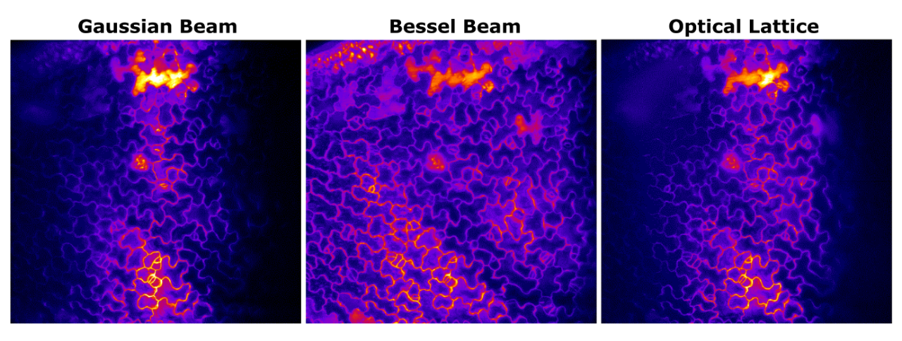
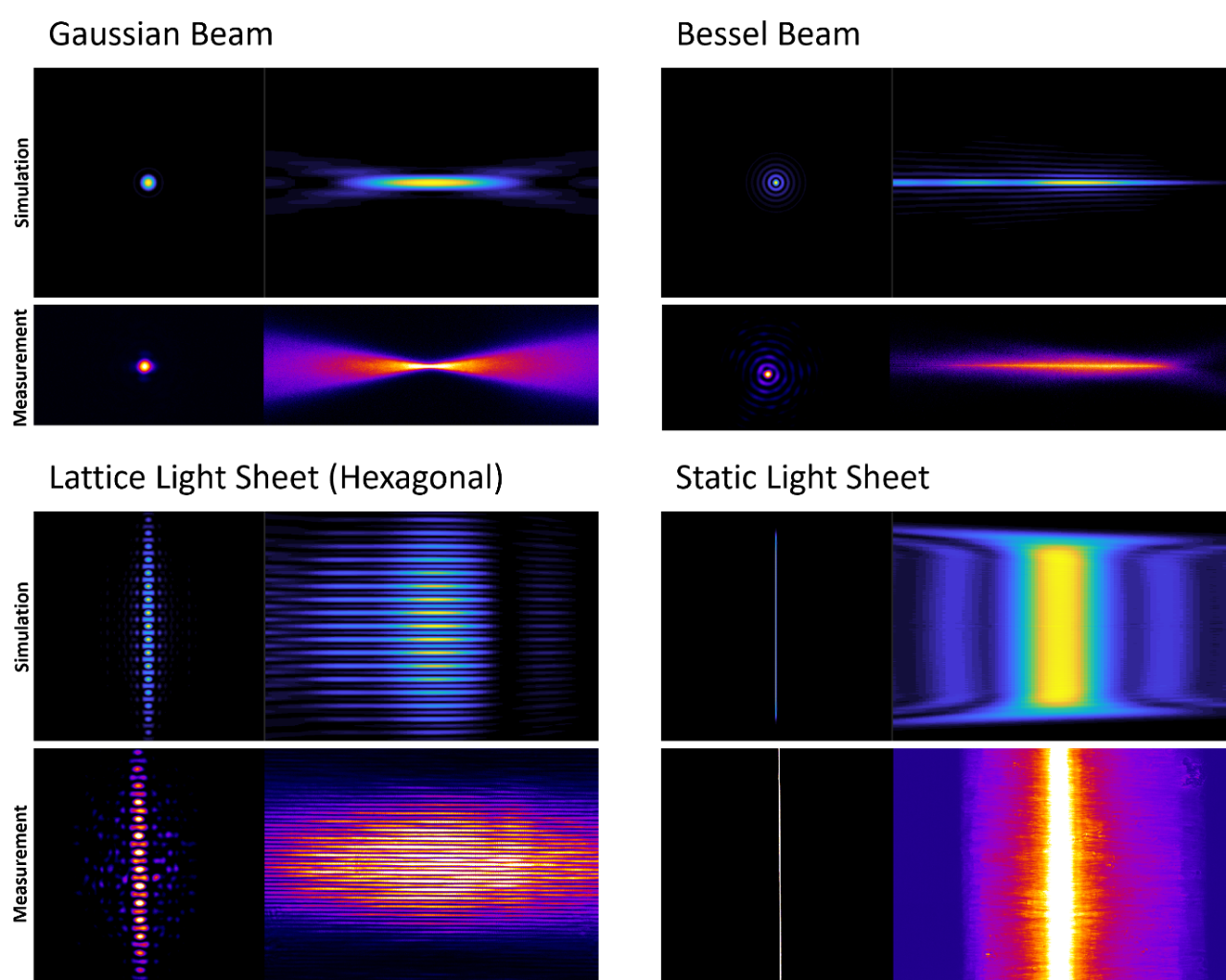
Every sample and application require diligently optimized experiment settings. The InVi SPIM Lattice Pro provides flexibility for fast adaptation of both illumination and detection to achieve the best results. A simple software command allows the system to switch between beam patterns such as Gaussian, Bessel, or Lattice Beams for multi-pattern illumination imaging in one experiment. Additionally, the Advanced Illumination Module (AIM) employs a fully integrated SLM to generate a broad range of illumination patterns featuring a characteristic pattern scanned vertically across the FOV to generate the light sheet.
The Gaussian beam has a waist at the center and diverges towards the periphery of the FOV. Scanning to generate a light sheet creates a small central area of the highest resolution covered by a thin light sheet. To overcome this limitation, the beam can be tuned for a more extended yet thicker waist, i.e., larger usable FOV at the price of overall resolution and contrast. Overall, a Gaussian beam is robust against scattering and well-suited for fast imaging. The classical static 2D Gaussian beam illumination provides all means for the highest speed continuous imaging. It is suitable for quantitative imaging modalities such as fluorescence lifetime imaging microscopy (FLIM) or fluorescence correlation spectroscopy (imaging FCS).
Self-reconstructing Bessel beams maintain a main lobe as thin as a Gaussian beam waist over a much more axial section than a Gaussian beam. This makes the Bessel beam the optimal choice for a large FOV at optimal resolution.
Optical lattice light sheets even out the Bessel beam side lobes through destructive interference, reducing light sheet thickness and non-specific fluorescence. The available lattice patterns have varying beam densities and characteristics along the different spatial dimensions, which affect resolution and FOV. Optical lattices can be used for resolution-enhancing structured illumination microscopy. Additionally, deconvolution can be performed during image processing to improve data quality further.
| Beam Pattern | Static Light Sheet | Scanned Gaussian | Scanned Bessel | Optical Lattices |
|---|---|---|---|---|
| Measured Resolution Z [1] | 800 nm | 750 nm | 680 nm | 530 nm |
| Measured Resolution XY [1] | 310 x 310 nm | 310 x 310 nm | 310 x 310 nm | 310 x 310 nm |
| Minimum LS thickness [2] | < 800 nm | < 800 nm | < 690 nm | < 550 nm |
| Max. FOV | 426 x 426 µm (31.25x) | 426 x 426 µm (31.25x) | 426 x 426 µm (31.25x) | 26.6 x 57 µm (62.5x) [3] |
| Acquisition Speed | 80 fps [4] | 80 fps [4] | 80 fps [4] | 80 fps [4] |
| Multi-Channel Imaging | simultaneous dual-color | simultaneous dual-color | simultaneous dual-color | sequential dual-color |
| Number of Preset Beam Patterns | 4 | 13 | 10 | 42 |
| Key Advantage | Highest-speed imaging and techniques such as FCS, FLIM | Beam robustness, fast imaging | Homogeneous illumination across a large FOV | Optimized and enhanced resolution in 3D |
Table depicting the specifications of different illumination patterns.
1 FWHM measured with 170 nm sized fluorescent beads / no deconvolution / @ 515 nm detection
2 Measured in water with 488 nm laser
3 Dithered light sheet
4 Full-frame imaging / max. 500 fps at reduced FOV
Advanced Imaging Methods
| Illumination |
|
| Beam Patterns |
|
| Beam Scanning Modes |
|
Next-Generation Structured Illumination Module (SIM)
Structured illumination microscopy (SIM) uses spatially structured patterns of excitation light to generate patterns of light interference, called the Moiré effect. When excitation pattern frequencies are correctly applied, SIM can result in a 50% lateral resolution enhancement.
Importantly, light is only maximally structured inside the focal plane. In contrast, out-of-focus light is homogenous enough to allow for its removal in image processing.
While no specific sample preparation is required, SIM requires longer exposure times and higher signal-to-noise ratios. While increased sub-diffraction resolution applies to the lateral resolution, the axial resolution is still diffraction-limited as the illumination pattern is not structured along the axial direction (Vangindertael et al. 2018 Methods Appl. Fluoresc. 6 022003).
Advantageous Photomanipulation
The ability to spatiotemporally control, manipulate, and alter biological processes is critical to revealing relevant mechanisms. A photomanipulation module (PM) can be added to InVi SPIM Lattice Pro, allowing for experiments such as photoablation, cauterization, optogenetics, and fluorescence recovery after photobleaching (FRAP) with:
- Beam coupled into detection objective
- Diffraction-limited illumination spot
- Flexible region-of-interest (ROI) generation (point, straight line, freeform, and square)
- Laser from continuous wave ultraviolet/visible (CW UV/VIS) to pulsed infrared (IR)
Full Environmental Control for Live Samples
While imaging living specimens, sample health and integrity are critical. The InVi SPIM Lattice Pro is equipped with an environmental control that supports long-term imaging (hours to days) of a wide variety of living specimens. This system is equipped with a generous sample chamber for immersion medium that controls temperature (20 to 37°C), gas concentration (CO2, O2, N2), and humidity.
Next-Level Resolution
The InVi SPIM Lattice Pro is the best solution for sample-preserving, long-term imaging at diffraction-limited resolution. Using two water-dippings and high NA objectives guarantees high acquisition quality while the adaptable magnification feature enables users to tailor the FOV to their needs. Additionally, the system's versatility is enhanced by its capacity for up to six solid-state laser lines, chromatic correction, a choice of 20 selectable filters, and dual-color acquisition, making it suitable for various applications.
Sample Mounting
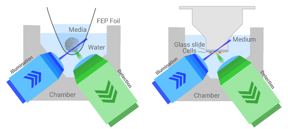
The InVi SPIM Lattice Pro has an inverted geometric design, easy sample, access, and accurate environmental control. Depending on beam patterns and experiments, sample mounting can be adapted to optimize resolution and experimental outcomes.
Broad Sample Variety
The InVi SPIM Lattice Pro is suitable for a variety of samples in cell biology from organoids and cell cultures to organs. Easy sample access allows straightforward sample placement and manipulation while switching beam patterns via a simple mouse click allows for experiment optimization.
Applications made possible with the InVi SPIM Lattice Pro include:
- Live samples' dynamic processes and interactions
- Cell cycle imaging
- Subcellular structure visualization
- Tracking of cells or protein movements in 3D
- Long, time-lapse imaging
- 2D cells, spheroid, organoid, tissue, or organism development
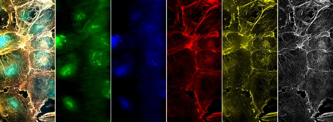
Subcellular Components
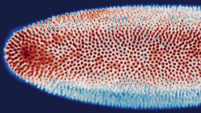
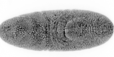
The InVi SPIM Lattice Pro is ideal for imaging subcellular components including cytoskeletal structures like actin.
The InVi SPIM Lattice Pro in Research
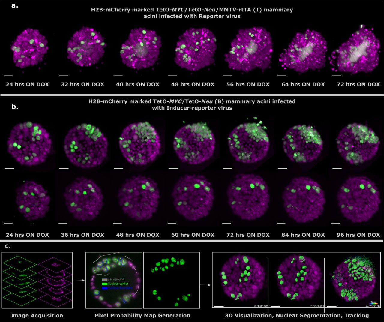
Primary mouse mammary epithelial acini to study oncogenes
Publication: Alladin et al. (2020), Tracking cells in epithelial acini by light-sheet microscopy reveals proximity effects in breast cancer initiation, eLife 9:e54066, DOI:10.7554/eLife.54066
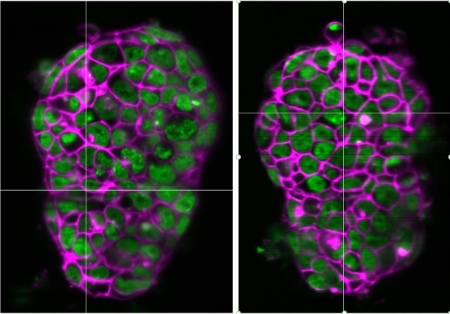
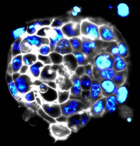
The InVi SPIM Lattice Pro is suitable for 3D Matrigel cultures of mammary acini. The imaging chamber allows for environmental control and has a reservoir for the immersion medium.
In their research work, Alladin et al. (2020) developed primary mouse mammary epithelial acini to study oncogenes.
To follow up their observations over time, they combined a nuclear reporter H2B-mCherry into the T and B mice to mark all the cells. The T/H2B-mCherry acini transduced with reporter virus proliferated upon doxycycline addition, showing an expansion of both the marked and unmarked cells. Time-lapse imaging showed tumor phenotype manifestation as seen by multi-cell-layered rims and pronounced proliferation-associated apoptosis in all acini.
Stem Cells
Pluripotent cells can self-renew and differentiate, with hundreds of genes expressed stably during self-renewal and directed expression upon signaling for differentiation. Characterizing the pluripotent state requires markers, such as miRNAs, which can also aid in differentiation. Research has shown that miRNA expression in human cells reveals surprising diversity and can be quantitatively mapped to understand stem cell processes. Colonies of mouse embryonic stem cells can be studied to realize pluripotency, involved genes, and regulatory networks.
Morphogenesis and Biophysics
The InVi SPIM Lattice Pro is an outstanding tool for visualizing developmental biology and morphogenesis with unprecedented detail. Taking this a step further, the optional photomanipulation module allows for:
- Control of fluorophores (optogenetics, uncaging, etc.)
- Study of dynamics (FRAP experiments)
- Biophysical studies (photoablation and cauterization)
Objective Specifications
| Objective | Magnification | Pixel Size | FOV |
|---|---|---|---|
| 25x 1.1NA | 31.3x | 208 nm | 426 µm |
| 62.5x | 104 nm | 213 µm |
System Comparison
* COMPATIBLE MODULE
| The perfect allrounder - multi-view imaging for diverse samples (live and cleared option) | Multi-sample and dual view flexibility for highly sensitive imaging, ideal for organoids and 3D cell cultures | In vivo multi-sample and multi-condition imaging, ideal for drug treatment comparison | Perfection for cleared samples prepared by any type of clearing protocol | High-end imaging with various beam patterns from single cells to 3D cell cultures | |
|---|---|---|---|---|---|
| Geometry | Horizontal with multiview | Horizontal with dual view | Inverted with dual illumination | Inverted dual illumination with moving optics | Inverted geometry at 360° |
| Beam Type | Gaussian | Gaussian and Bessel | Gaussian | Gaussian | Advanced Illumination Module (AIM) |
| Application Examples | - Long-term imaging of Drosophila development - Photomanipulation studies in zebrafish embryos - Cleared and stained samples up to mouse brain size | - High-resolution full 3D imaging of multiple organoids over time - Long-term imaging of highly light-sensitive samples - Comparing the pharmacological impact in zebrafish wound healing after PM | - Time-lapse imaging of multiple pancreatic spheres - Studying pharmacological impact on zebrafish development - Comparing genetically altered organoids | - Pharmacokinetics in mouse brain - Multi-stained rat brain - Whole mouse imaging | - Fast imaging of photo-sensitive 2D cell culture - Lattice imaging for enhanced resolution in 3D cell cultures - FCS and FLIM in cell culture |
| User Level | Beginner friendly | Beginner friendly | Beginner friendly | Intermediate | Advanced |
| # Lenses | 4 lenses (2 IO, 2 DO) | 3 lenses (1 IO, 2 DO) | 3 lenses (2 IO, 1 DO) | 3 lenses (2 IO, 1 DO) | 2 lenses (1 IO, 1 DO) |
| Multi-View | ✓ | ✓ | |||
| Live/Fixed Samples | ✓ | ✓ | ✓ | ✓ | |
| Cleared Samples | ✓ | ✓ | |||
| Expanded Samples | ✓ | ✓ | ✓ | ✓ | ✓ |
| Best Embedding | Capillary/FEP tube w/ agarose 3D stage | Custom-solution and FEP foil | TruLive3D dishes and FEP foil | Quartz-crystal cuvette | FEP foil; glass slides |
| Photomanipulation* | ✓ | ✓ | ✓ | ✓ | |
| Environmental Control* | ✓ | ✓ | ✓ | ✓ | |
| Destriping/Uniform Illumination* | ✓ | ✓ | ✓ | ✓ | ✓ (w/ Advanced Illumination Module) |
| Benchtop Design | ✓ | ✓ | ✓ | ✓ | ✓ |
All-in-One LuxBundle Software
Intuitive design
Bruker's LuxBundle software saves time and enhances productivity by providing:
- All-in-one, easy-to-use interface for acquisition, viewing, and post-processing
- Fully scriptable microscope control and post-processing via open interface (e.g., Python or any other language), ready for custom "smart" microscopy
- High reproducibility of experiments: all parameters are saved in the metadata and configurations can be saved for future experiments
- Data formats (.tiff, .hdf5, .ims) compatible with common image processing software: Imaris, Aivia, BigDataViewer, Arivis, Fiji, Python, Matlab, Napari
Impressive Image Post Processor
Our dual-view light-sheet microscopes record a sample from different angles/views and generate images composed of multiple tiles. The LuxBundle software ensures high-quality, 360° crisp images of the sample that compensate for absorption and scattering. Features include:
- Multi-color alignment
- Tile stitching of hundreds of tiles for large samples
- Multi-view image fusion and deconvolution
3D Data Viewer
LuxBundle's integrated 3D data viewer allows researchers to inspect the entire dataset directly after acquisition. This gives users control over their data with key capabilities, including:
- The ability to turn tile stitching on or off
- Both raw and post-processed images
- Fast viewing of multi-terabyte data sets
- Flexible options to draw and annotate regions and landmarks