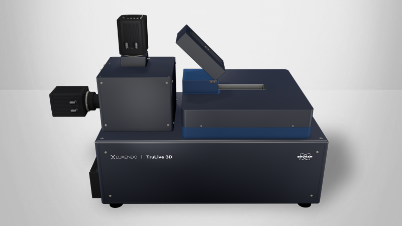
Light-Sheet Microscopes
See Biology Across All Scales
With a strong focus on applications, our light-sheet solutions are easy to use and enable scientists around the globe to excel in their research. The SPIM technology avoids sample phototoxicity by sequentially illuminating a stack of thin slices of the organism, allowing scientists to observe living organisms for extended periods of time without photodamage.

System Comparison
* COMPATIBLE MODULE
| The perfect allrounder - multi-view imaging for diverse samples (live and cleared option) | Multi-sample and dual view flexibility for highly sensitive imaging, ideal for organoids and 3D cell cultures | In vivo multi-sample and multi-condition imaging, ideal for drug treatment comparison | Perfection for cleared samples prepared by any type of clearing protocol | High-end imaging with various beam patterns from single cells to 3D cell cultures | |
|---|---|---|---|---|---|
| Geometry | Horizontal with multiview | Horizontal with dual view | Inverted with dual illumination | Inverted dual illumination with moving optics | Inverted geometry at 360° |
| Beam Type | Gaussian | Gaussian and Bessel | Gaussian | Gaussian | Advanced Illumination Module (AIM) |
| Application Examples | - Long-term imaging of Drosophila development - Photomanipulation studies in zebrafish embryos - Cleared and stained samples up to mouse brain size | - High-resolution full 3D imaging of multiple organoids over time - Long-term imaging of highly light-sensitive samples - Comparing the pharmacological impact in zebrafish wound healing after PM | - Time-lapse imaging of multiple pancreatic spheres - Studying pharmacological impact on zebrafish development - Comparing genetically altered organoids | - Pharmacokinetics in mouse brain - Multi-stained rat brain - Whole mouse imaging | - Fast imaging of photo-sensitive 2D cell culture - Lattice imaging for enhanced resolution in 3D cell cultures - FCS and FLIM in cell culture |
| User Level | Beginner friendly | Beginner friendly | Beginner friendly | Intermediate | Advanced |
| # Lenses | 4 lenses (2 IO, 2 DO) | 3 lenses (1 IO, 2 DO) | 3 lenses (2 IO, 1 DO) | 3 lenses (2 IO, 1 DO) | 2 lenses (1 IO, 1 DO) |
| Multi-View | ✓ | ✓ | |||
| Live/Fixed Samples | ✓ | ✓ | ✓ | ✓ | |
| Cleared Samples | ✓ | ✓ | |||
| Expanded Samples | ✓ | ✓ | ✓ | ✓ | ✓ |
| Best Embedding | Capillary/FEP tube w/ agarose 3D stage | Custom-solution and FEP foil | TruLive3D dishes and FEP foil | Quartz-crystal cuvette | FEP foil; glass slides |
| Photomanipulation* | ✓ | ✓ | ✓ | ✓ | |
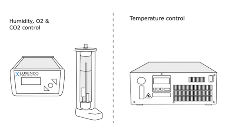
| Environmental Control* | ✓ | ✓ | ✓ | ✓ | |
| Destriping/Uniform Illumination* | ✓ | ✓ | ✓ | ✓ | ✓ (w/ Advanced Illumination Module) |
| Benchtop Design | ✓ | ✓ | ✓ | ✓ | ✓ |


Watch Our Light-Sheet Microscopy Webinars
Our webinars cover best practices, introduce new products, provide quick solutions to tricky questions, and offer ideas for new applications, modes, or techniques.
Browse Light-Sheet Microscopy Articles
| Year | Journal | Title | Author(s) | Imaging Platform | Subject | Methodology |
|---|---|---|---|---|---|---|
| 2026 | BMC Bioinformatics | LimbLab: Pipeline for 3D Analysis and Visualisation of Limb Bud Gene Expression | Laura Aviñó-Esteban, Heura Cardona-Blaya, Marco Musy, Antoni Matyjaszkiewicz, James Sharpe, Giovanni Dalmasso | MuVi SPIM | morphogenesis, limb development, biomedical image analysis | Python, 3D Visualization, Limb Development, Gene Expression Analysis, Bioinformatics Pipeline |
| 2026 | Cell Stem Cell | 3D post-implantation co-culture of human embryo and endometrium | Song et al | MuVi SPIM | endometrium, embryo-maternal interaction, co-culture, development | 3D co-culture system, human, organoids, immunostaining, sequencing |
| 2026 | Biorxiv preprint | 3D Light Sheet Microscopy Reveals Aging Osteoarthritis-associated Joint-innervated Nerve Remodeling in Mouse Knee Joints | Paromita Kundu, Brian Wise, Emma Norris, Mark James, Yaxin Zhang, Jennifer Jonason, Mark Buckley, Whasil Lee | MuVi SPIM | Aging osteoarthritis, joint pain, joint-innervating nerve, Dorsal root ganglion | mice, 3D Light-Sheet imaging, iDISCO clearing, image analysis |
| 2026 | eLife | Glial betaPix is essential for blood vessel integrity in the zebrafish brain | ShihChing Chiu, Qinchao Zhou, Chenglu Xiao, Linlu Bai, Xiaojun Zhu, Wanqiu Ding, Jing-Wei Xiong | MuVi SPIM | betaPix, zebrafish, brain | zebrafish, cell culture, transfection, mRNA/gRNA synthesis and microinjections, knockout, drug treatment, WISH |
| 2025 | Periductal fibroblasts participate in liver homeostasis, fibrosis, and tumorigenesis | Shan-Shan Wang, Jia Yuan, Xinyu Thomas Tang, Xiujuan Yin, Ke Fang, Lin Veronica Chen, Zhenggang Ren, Bo O. Zhou | MuVi SPIM | fibroblast, liver, fibrosis, cancer | ||
| 2025 | eLife | Tgfbr1 regulates lateral plate mesoderm and endoderm reorganization during the trunk to tail transition | Anastasiia Lozovska, Ana Casaca, Ana Novoa, Ying-Yi Kuo, Arnon D Jurberg, Gabriel G Martins, Anna-Katerina Hadjantonakis, Moises Mallo | MuVi SPIM | Tgfbr1, isl1, LPM, trunk-to-tail transition, endoderm, mesoderm | mouse, Whole-mount in situ hybridization, immunofluorescence, Ex vivo culture |
| 2025 | Biorxiv preprint | L1CAM signaling through planar cell polarity generates SOX2+ metastatic progenitors in lung adenocarcinoma | Jin Suk Park, Yasemin Kaygusuz, Carson Kenum, Lan He, Mohamed I. Gatie, Xueqian Zhuang, Roshan Sharma, Ronan Chaligné, Umesh K. Bhanot, Richard P. Koche, Tuomas Tammela, Anna-Katerina Hadjantonakis, Charles M. Rudin, Joan Massagué | MuVi SPIM | lung, adenocarcinoma, cancer, L1CAM, WNT | mice, tumor initiation, Micro-CT imaging, clearing, xenografts |
| 2025 | The EMBO Journal | Rbm24a dictates mRNA recruitment for germ granule assembly in zebrafish | Yizhuang Zhang, Jiasheng Wang, Hailing Fang, Shuqi Hu, Boya Yang, Jiayi Zhou, Raphaëlle Grifone, Panfeng Li, Tong Lu, Zhengyang Wang, Chong Zhang, Yubin Huang, Dalei Wu, Qianqian Gong, De-Li Shi, Ang Li & Ming Shao | MuVi SPIM | primordial germ cells, rbm24a, germ granules, RNA | zebrafish, knockout, immunostaining |
| 2025 | Biorxiv preprint | A scalable human-zebrafish xenotransplantation model reveals gastrosome-mediated processing of dying neurons by human microglia | Ambra Villani, Jana Wittmann, Tamara Wyss, Izaskun Mallona Gonzalez, Irene Santisteban Ortiz, Nathalie Tichy, Monique Pena, Ayush Aditya Pal, Darren Gilmour, Simon T. Schafer and Francesca Peri | TruLive3D Imager | xenotransplantation, microglia, Niemann-Pick disease | zebrafish, microscopy, Microglia |
| 2025 | Cell | Live imaging endogenous transcription factor dynamics reveals mechanisms of epiblast and primitive endoderm fate segregation | Rebecca P. Kim-Yip, David Denberg, Denis F. Faerberg, Hayden Nunley, Isabella Leite, Madeleine Chalifoux, Bradley Joyce, Jared Toettcher, Bin Gu, Eszter Posfai | InVi SPIM | epiblast, endoderm, development, NANOG, EPI/PE segregation | Live image acquisition, image analysis, Immunofluorescence, mice |
| 2025 | eBioMedicine | The effect of amycretin, a unimolecular glucagon-like peptide-1 and amylin receptor agonist, on body weight and metabolic dysfunction in mice and rats | Rune Ehrenreich Kuhre et al | Glucagon-like peptide-1 receptor; Amylin receptor; Metabolism; Obesity | mice, rats, In vitro assessment | |
| 2025 | Biorxiv preprint | A mechanical origin for implantation defects in embryos from aged females | Kate E. Cavanaugh, MJ Franco-Oñate, Diana J. Laird, Patrick W. Oakes, Ricard Alert, Orion D. Weiner | TruLive3D Imager | fertility, biomechanics, implantation | Traction Force Microscopy, Live imaging, Cell shape analysis, Immunofluorescence, NSET Procedures. |
| 2025 | ResearchSquare preprint | Phage–antibiotic synergy restores β-lactam efficacy in MDR Klebsiella quasipneumoniae biofilms and suppresses resistance | Tinatini Tchatchiashvili, Mike Marquet, Ekaterine Gabashvili, Kamran A. Mirza, Mara Lohde, Christian Brandt, Ralf Ehricht, Mathias W. Pletz, Oliwia Makarewicz | TruLive3D Imager | Biofilms, phage–antibiotic synergy | Biofilm, drug treatment |
| 2025 | Biorxiv preprint | Automated cell naming reveals reproducible and variable features of ascidian embryogenesis | Kilian Biasuz, Haydar Jammoul, Benjamin Gallean, YanisAsloudj, Julien Laussu, Patrick Lemaire and Gregoire Malandain | MuVi SPIM | cell lineage, modelling | Phallusia mammillata, Ascidiella aspersa, biomedical image analysis, |
| 2025 | Biorxiv preprint | Actinotrichia-independent developmental mechanisms of spiny rays facilitate the morphological diversification of Acanthomorpha fish fins | Kazuhide Miyamoto, Junpei Kuroda, Satomi Kamimura, Yasuyuki Sasano, Gembu Abe, Satoshi Ansai, Noriko Funayama, Masahiro Uesaka, Koji Tamura | QuVi SPIM | morphological diversification, extracellular matrix, cell biology | Melanotaenia praecox, Stephanolepis cirrhifer, X-ray micro-CT scanning, DAR4M and SYTO13 staining |
| 2025 | Biorxiv preprint | Mesoglea biogenesis reveals a cryptic aboral valve for pressure regulation in cnidarian morphogenesis | Soham Basu, Petrus Steenbergen, Florian Gabler, Alexandre Paix, Paolo Ronchi, Gleb Bourenkov, Thomas Schneider, Jonas Hellgoth, Anna Kreshuk, Suat Özbek, Aissam Ikmi | MuVi SPIM | multicellular organisms, development, biomechanics | Cnidarians, Live imaging, Photoconversion |
| 2025 | Journal of Advanced Research | Glycyrrhetinic acid augments lipid catabolism via immune-neural modulation in adipose tissue | Wenjiao Jiang, Sasa Zhang, Xinyuan Sun, Shun Wang, Jinwei Zhu, Ziao Liu, Wanting Zhang, Huijie Guo, Hanwen Li, Hao Xie, Kun Hao | MuVi SPIM | metabolism, neural modulation, adipose tissue, macrophages | mice, histopathology, metabolic monitoring, drug treatment, immunoprecipitation |
| 2025 | Biorxiv preprint | Mechanics and growth coordination define SOSEKI-based polarity fields | Marcel Piepers, Blanca Jazmin Reyes-Hernández, João R D Ramos, Neva Bölke, Laura Schütz, Changzheng Song, Anamarija Primc, Lotte Bald, Roeland MH Merks, Dolf Weijers, Alexis Maizel | MuVi SPIM | biomechanics, plant development | Live imaging, Arabidopsis thaliana, Laser ablation |
| 2025 | Biological and Medical Applications of Materials and Interfaces | Fluorescent Molecular Probe for Imaging Hypoxia in 2D Cell Culture Monolayers and 3D Tumor Spheroids: The Cell Membrane Partition Model for Predicting Probe Distribution in a Spheroid | Zhumin Zhang, Hailey S. Sanders, Vivienne Dragun, Sara Cole, Bradley D. Smith | MuVi SPIM | Cells, Fluorescence, Membranes, Probes, Tumors | tumor spheroid, fluorescence microscopy, hypoxia, nitroreductase, membrane partition, coefficient |
| 2025 | Biorxiv preprint | 3D Mapping of Intact Ovaries Reveals the Aging Dynamics of the Ovarian Reserve | Arturo D’Angelo, Daniel Franco-Barranco, Marco Musy, James Sharpe, Ignacio Arganda-Carreras, Elvan Böke | MuVi SPIM | oocytes, fertility, organ-scale regulatory mechanism | immunostaining, BABB clearing, tissue clearing |
| 2025 | Angiogenesis | Cxcl9 modulates aging associated microvascular metabolic and angiogenic dysfunctions in subcutaneous adipose tissue | Xin Fu, Yu Zhao, Xiwei Cui, Siyuan Huang, Yanze Lv, Chen Li, Fuxing Gong, Zhigang Yang, Xiaonan Yang & Ran Xiao | LCS SPIM | Microvascular aging, Endothelial cells, Metabolism, Cxcl9, Aplnr, Angiogenesis | cell culture, 3D volume fluorescence, immunohistochemistry, mouse |
| 2025 | Nature | Patterned invagination prevents mechanical instability during gastrulation | Bruno C. Vellutini, Marina B. Cuenca, Abhijeet Krishna, Alicja Szałapak, Carl D. Modes & Pavel Tomancak | MuVi SPIM | cephalic furrow | Drosophila, epithelial fold, biomechanics, in vivo perturbations and in silico simulations |
| 2025 | Developmental Biology | Geometric, cell cycle and maternal-to-zygotic transition-associated YAP dynamics during preimplantation embryo development | Madeleine Chalifoux, Maria Avdeeva, Eszter Posfai | InVi SPIM | YAP, CDX2, SOX2, ICM cells, embryogenesis | mouse, Image analysis, live imaging, |
| 2025 | Anatomical Science International | ht-MASH: a high-throughput, cost-effective, and robust protocol for microscopic 3D imaging of human angio-and cytoarchitecture in large human brain samples | Sven Hildebrand, Johannes Franz, Shubharthi Sengupta, Anna Schueth, Andreas Herrler & Alard Roebroeck | QuVi SPIM | human brain tissue, 3D printing, tissue clearing | tissue clearing, labelling, cytoMASH , |
| 2025 | Jove | Precision Ultrasound-guided Stem Cell Delivery for Vascular Repair in Aortic Diseases | Qiuyue Gao, Qing Xu, Xuejie Cao, Xian Xu, Qingbo Xu, Chen Zhang, Baoqi Yu | MuVi SPIM | ultrasound-guided delivery, vascular biology, aneurysm | |
| 2025 | Biorxiv preprint | Multiscale wrinkling dynamics in epithelial shells | Nimesh Chahare, Adam Ouzeri, Thomas Wilson, Pradeep K. Bal, Tom Golde, Guillermo Vilanova, Pau Pujol-Vives, Pere Roca-Cusachs, Xavier Trepat, and Marino Arroyo | QuVi SPIM | cytoskeleton, epithelium, cell buckling, biomechanics, cortex | 3D computational model, microfluidic devices, cell culture |
| 2025 | Biorxiv preprint | Deep Tissue Second-Harmonics of Collagen Fibers in a Transparent Rat Heart from a Myocardial Infarction Model | Makoto Matsuyama | MuVi SPIM, LCS SPIM | rodent model of myocardial infarction, whole heart transparency, second-harmonics | tissue-clearing, label-free imagin, rat |
| 2025 | Springer Nature | Live Imaging of Mouse Embryonic Stem Cells and Embryos: Protocols for In Vitro Culture, Microscopy, and Image Data Analysis | Woonyung Hur, Mohamed I. Gatie, Alexandre Francou & Anna-Katerina Hadjantonakis | TruLive 3D Imager | protocol, reproduciblity, embryo imaging compatibility | mouse, pluripotent stem cells |
| 2025 | STAR Protocols | Protocol for analyzing T cell and macrophage motility in a mouse pancreatic cancer spheroid model using light-sheet microscopy | Vanessa Zimmer, Benedikt Slusny, Katrin Roth, Christian Bauer | TruLive 3D Imager | cell culture, immunology, microscopy, spheroid | spheroid, co-culturing, mouse |
| 2025 | Journal of Mammary Gland Biology and Neoplasia | Live-cell imaging of mammary organoids using light sheet microscopy | Matea Brezak, Azqa Ajmal Khan, Martin Jechlinger & Zuzana Sumbalova Koledova | TruLive 3D Imager | tissue culture, imaging, organoids | dissection, organoid, multiview LSM |
| 2025 | Cell | Post-gastrulation amnioids as a stem cell-derived model of human extra-embryonic development | Borzo Gharibi, Oliver C.K. Inge, Irene Rodriguez-Hernandez, Paul C. Driscoll, Christelle Dubois, Ming Jiang, Michael Howell, J. Mark Skehel, James I. Macrae, Silvia D.M. Santos | MuVi SPIM | Post-gastrulation amnioid (PGA), GATA3 , amniogenesis, BMP, Ent | CRISPR–Cas9, human ES, 3D extra-embryonic PGA model, Immunofluorescence |
| 2025 | Princeton University Thesis | YAP Regulation in the Preimplantation Embryo and Its Role in the First Cell Fate Decision | Chalifoux, Madeleine | InVi SPIM | YAP, CDX2, SOX2, ICM cells, embryogenesis | mouse, Image analysis, live imaging, |
| 2025 | Nature Structural & Molecular Biology | Maternal ELL3 loss-of-function leads to oocyte aneuploidy and early miscarriage | Shiqi Zhu, Peng Xie, Yi Yang, Yan Wang, Chuanxin Zhang, Yu Zhang, Shuhan Si, Jin Zhang, Jingjing He, Hao Si, Ke Fang, Binbin Ma, Xu Jiang, Lindi Huang, Jiamin Li, Tian Min, Beihong Zheng, Lincui Da, Dianliang Lin, Kun Gao, Yuanyuan Li, Mingtao Huang, Fengchang Qiao, Haiqin Huo, …Chengqi Lin | InVi SPIM | aneuploidy, miscarriage, development, meiotic spindle, meiosis | |
| 2025 | Biorxiv preprint | The synovial lining macrophage layer develops in the first weeks of life in a CSF1-and TGFβ-dependent but monocyte-independent process. | Marlene Magalhaes Pinto, Bert Malengier-Devlies, Guillaume Seuzaret, Anna Ahlback, Solvig Becker, Katelyn Patatsos, Georgios Drakoulis, Julia Karjalainen, ChrisNane Ruedl, ... Rebecca Gentek | MuVi SPIM | Synovial joints, fibroblasts, macrophages, development | scRNAseq analysis, RNA sequencing, mouse, drug treatments, |
| 2025 | Biorxiv preprint | High-Quality Cell Capture Reveals Transcriptomic Changes After Single-Axon Injury of Mauthner Cells | Zheng Song, Lin Zhu, Yueru Shen, Huaitong Yao, Yuan Cai, Lingyu Shi, Xinghan Chen, Along Han, Ziang Zhao, Kun Qu, Bing Hu | MuVi SPIM | Mauthner Cells, axon injury, transcriptomics | Two-photon axotomy, Light-sheet microscope imaging, Library construction, transcriptome sequencing, cell scoring, |
| 2025 | Reviews in Fish Biology and Fisheries | Ontogenetic and evolutionary trends on cephalopod digestive systems | Diego G. Vilarnau, Fernando Á. Fernández-Álvarez, Montserrat Coll-Lladó, Nicola Gritti, Jim Swoger & Roger Villanueva | LCS SPIM, MuVi SPIM | digestive systems, biological niche, morphogenesis, ontogeny | octopuses, image analysis, |
| 2025 | Nature Communications | Nuclear position controls the activity of cortical actomyosin networks powering simultaneous morphogenetic events | Nicolas Roby & Matteo Rauzi | MuVi SPIM | gastrulation, nuclear positioning, morphogenesis, RhoGEF2 | Drosophila, live imaging, In toto 4D imaging, laser dissection, Optogenetic recruitment, In silico 3D modelling |
| 2025 | Ecotoxicology and Environmental Safety | Neomycin affects cardiovascular and hematopoietic system via the PI3K/Akt pathway in zebrafish larvae | Yuan Lin, Qiuping Zhang, Lu Chen, Yingying Liu, Xiaoxi Lin, Xiaoyan Peng, Hua Cao, Yuqing Lei, Xinrui Wang | MuVi SPIM | PI3K/Akt, neomycin, toxicology, hematopoietic system | zebrafish, Toxicity tests, Behavior assay, genetic evaluations, biochemistry, Neutrophil, macrophage, ROS |
| 2025 | Biorxiv preprint | Generative model for the first cell fate bifurcation in mammalian development | Maria Avdeeva, Madeleine Chalifoux, Bradley Joyce, Stanislav Y. Shvartsman, Eszter Posfai | InVi SPIM | cell fate bifurcation, YAP, CDX2, SOX2, ICM cells, embryogenesis | mouse, Image analysis, live imaging, |
| 2025 | Biorxiv preprint | Boundary-guided cell alignment drives mouse epiblast maturation | Takafumi Ichikawa, Pamela C. Guruciaga, Shuchang Hu, Steffen Plunder, Mei Makino, Marina Hamaji, Anniek Stokkermans, Shinjiro Yoshida, Anna Erzberger, Takashi Hiiragi | InVi SPIM | 3D-geec embryos, symmetry breaking, non-polarized epiblast (EPI) cells orientation dynamics, ERK activation | mouse, 3D-gel embedded embryo culture (3D-geec), Immunofluorescence, Light-sheet Live-imaging, Simulations, Image analysis, machine-learning |
| 2025 | American Journal of Cell Physiology | Comprehensive evaluation and application of tissue clearing techniques for 3-D visualization of splenic neural and immune architecture | Jianing Li*,Letian He*,Wenling Wang*,Siyu Wang,Dan Zhang,Liyun Liang,Guangping Song,Yijian Zhang,Shaoqing Yu,Lei Wang,Qiuying Han,Shaoyi Huang,Sen Li,Haiqing Tu,Zengqing Song,Huaibin Hu,Huiyan Li,Yang Yang, andMin Wu | MuVi SPIM | spleen, immune response, immune and neural components, | tissue clearing, ImmuView, FLASH, Ce3D, SHANEL, advanced CUBIC |
| 2025 | Pulmonary Circulation | Multiscale Three‐Dimensional Evaluation and Analysis of Murine Lung Vasculature From Macro‐to Micro‐Structural Level | Birger Tielemans, Nora F. Marain, Axelle Kerstens, Nicolas Peredo, Montserrat Coll-Lladó, Nicola Gritti, Perrine de Villemagne, Paul Dorval, Vincent Geudens, Michaela Orlitová, Sebastian Munck, Bartosz Leszczyński, Jim Swoger, Greetje Vande Velde | LCS SPIM, MuVi SPIM | vasculature, analysis, experimental design, pulmonary vasculature | mouse, whole-organ view |
| 2025 | BJP | Deep learning‐enhanced 3D imaging unveils semaglutide impact on cardiac fibrosis | Sheyla Barrado-Ballestero, Sarah Torp Yttergren, Max Hahn, Marie Biviano Rosenkilde, Ditte Marie Jensen, Michael Christensen, Louise Thisted, Heidi Lindgreen Holmberg, Geoffrey Teixeira, Tor Biering-Sørensen, Casper Gravesen Salinas, Urmas Roostalu | LCS SPIM | fibrosis, image analysis, experimental design, cardiovascular diseases | deep learning, tissue clearing, |
| 2025 | Biorxiv preprint | Using lightsheet microscopy to investigate the initial lymphatic network in the murine knee joints | L. Xing, X. Lin | MuVi SPIM | established lightsheet imaging protocol | mouse, immunostaining |
| 2025 | Cancers | Optimized Spheroid Model of Pancreatic Cancer Demonstrates Influence of Macrophage–T Cell Interaction for Intratumoral T Cell Motility | Benedikt Slusny, Vanessa Zimmer, Elena Nasiri, Veronika Lutz, Magdalena Huber, Malte Buchholz, Thomas M. Gress, Katrin Roth, and Christian Bauer | TruLive 3D Imager | spheroid, Pancreatic Cancer, T cell motility | spheroid, Mouse, |
| 2025 | JCB | KIF2A maintains cytokinesis in mouse embryonic stem cells by stabilising intercellular bridge microtubules | Stockmann et al | InVi SPIM | cytokinesis, cell division, KIF2A | cell culture, antibodies, transgenic mESC lines, immunofluorescence, imaging, image analysis |
| 2024 | Ecotoxicology and Environmental Safety | Cardiovascular developmental hazards of valproic acid in zebrafish | Yuqing Lei, Yingying Liu, Wenpeng Xie, Yalan Wei, Xudong Zhuang, Haitao Zhang, Hua Cao, Xinrui Wang | MuVi SPIM | Valproic acid (VPA), embryology, development, toxicology | zebrafish, reactive oxygen species, apopotosis |
| 2024 | Life Medicine | 4D live tracing reveals distinct movement trajectories of meiotic chromosomes | Peng Xie , Shiqi Zhu , Jin Zhang , Xinrui Wang , Xu Jiang , Feng Xiong , Linjin Chen , Ke Fang , Yuanhui Ji , Beihong Zheng , Lincui Da , Hua Cao , Yan Sun , Zhuojuan Luo , Chengqi Lin | InVi SPIM | chromosome segregation, cell division, KIFs | deep learning, mouse, microinjections, Immunofluorescence, live cell imaging, |
| 2024 | Scientific Reports | Tumor biomechanics as a novel imaging biomarker to assess response to immunotherapy in a murine glioma model | Yannik Streibel, Michael O. Breckwoldt, Jessica Hunger, Chenchen Pan, Manuel Fischer, Verena Turco, Berin Boztepe, Hannah Fels-Palesandro, Jonas G. Scheck, Volker Sturm, Kianush Karimian-Jazi, Dennis A. Agardy, Giacomo Annio, Rami Mustapha, Shreya S. Soni, Abdulrahman Alasa, Ina Weidenfeld, Christopher B. Rodell, Wolfgang Wick, Sabine Heiland, Frank Winkler, Michael Platten, Martin Bendszus, Ralph Sinkus & Katharina Schregel | LCS SPIM | murine glioma, immunotherapy , myeloid influx, inflammation, Glioblastoma | magnetic resonance elastography, MRI, tissue clearing, immunohistochemistry, image analysis |
| 2024 | Cell Press | A mechanical wave travels along a genetic guide to drive the formation of an epithelial furrow during Drosophila gastrulation | A Popkova, U Andrenšek, S Pagnotta, P Ziherl, M Krajnc, M Rauzi | MuVi SPIM | morhphogenesis, epithelial furrowing, pacemakers | Drosophila, imaging, cytoskeleton |
| 2024 | Science | Temporal variability and cell mechanics control robustness in mammalian embryogenesis | D Fabrèges, B Corominas-Murtra, P Moghe, A Kickuth, (..) Takashi Hiiragi | InVi SPIM | Pre-implantaion development, embryogenesis, Temporal variability and cell mechanics, | mouse, rabbit and monkey; immunostaining, chemical inhibitors, in silico modelling, image analysis |
| 2024 | Communications Medicine | Structural reconstruction of mouse acute aortic dissection by intravenously administered human Muse cells without immunosuppression | M Takahashi, Y Kushida, Y Kuroda, S Wakao, Y Horibata, H Sugimoto, M Dezawa & Y Saiki | MuVi SPIM | Stanford type B acute aortic dissection (Stanford-B AAD), Muse cells | Muse cells, acute aortic dissection, Stanford type B, intravenous injection, sphingosine-1-phosphate, elastin, HLA-G |
| 2024 | RNA | Boosting the toolbox for live imaging of translation | M Bellec, R Chen, J Dhayni, (..) Jeremy Dufourt | MuVi SPIM | Live imaging of translation | drosophila, mammalian cells, nanobody/tag system, ALFA_array |
| 2024 | Ecotoxicology and Environmental Safety | Effects of water fluoridation on early embryonic development of zebrafish | Y-L Wei, X-C Lin, Y-Y Liu, Y-Q Lei, X-D Zhuang, H-T Zhang, X-R Wang | MuVi SPIM | fluoridation, environmental toxicology, embryogenesis, development | drug assay, LD50, zebrafish |
| 2024 | Nature Methods | Image restoration of degraded time-lapse microscopy data mediated by near-infrared imaging | N Gritti, R M. Power, A Graves & J Huisken | MuVi SPIM | time-lapse microscopy, image degradation, image restoration, AI, machine learning, neural network | AI, machine learning, neural network, near infra-red imaging |
| 2024 | Scientific Reports | Visualisation of gene expression within the context of tissues using an X-ray computed tomography-based multimodal approach | K Kairišs, N Sokolova, L Zilova, C Schlagheck, R Reinhardt, T Baumbach, T Faragó, T van de Kamp, J Wittbrodt & V Weinhardt | MuVi SPIM | multimodal imaging and registration, gene expression visualization, medaka | X-ray CT, light sheet microscopy, multimodal data |
| 2024 | JMRI | Assessment of Tumor Cell Invasion and Radiotherapy Response in Experimental Glioma by Magnetic Resonance Elastography | Fels-Palesandro et al. | LCS SPIM | glioma, MRI, tissue clearing | multimodal data, coregistration |
| 2024 | ResearchSquare preprint | Cross-species transcriptome analysis of the subthalamic and para-subthalamic nuclei reveal mRNA patterns unique to primate and relevant to neuropsychiatry | S Dumas, A Ricci, E Rubino, M Rosenkilde, C Francois, C Karachi, Å Wallen-Mackenzie | LCS SPIM | mRNA patterns, neuropsychiatry, brain regionalization | mRNA patterns, primate, mice, tissue clearing |
| 2024 | Front. Immunol. | Impaired meningeal lymphatic drainage in Listeria monocytogenes infection | J Feng, Y Ren, X Wang, X Li, X Zhu, B Zhang, Q Zhao, X Sun, X Tian, H Liu, F Dong, X-L Li, L Qi, and B Wei | MuVi SPIM | Listeria monocytogenes, bacterial infection, cellular polarization, inflammation, lymphangiogenesis | Mice, rtPCR, HE staining, tissue clearing, cytometry |
| 2024 | Development | Nuclear instance segmentation and tracking for preimplantation mouse embryos | Nunley et al. | |||
| 2024 | Thesis Harvard | Use of the Zebrafish to Explore the Connection of Pituitary Development with Congenital Craniofacial Syndromes | Tsay | craniofacial malformations, Esrp1/2, lhx3, embryogenesis | zebrafish, WISH, RNAscope, Crispr/Cas9, morpholino | |
| 2024 | Scientific Reports | Tumor biomechanics as a novel imaging biomarker to assess response to immunotherapy in a murine glioma model | Streibel et al. | LCS SPIM | MRI, MRE, glioblastoma, glioma | Multiparametric MRI and MRE, Tissue clearing and LSM, immunohistochemistry, image analysis |
| 2024 | biorxiv preprint | Creased ciliary flocks shape unfolding dynamics via information bottlenecks in an aneural animal | C Brannon and M Prakash | TruLiv3D Imager | Placozoa, Trichoplax adhaerens, epithelial folding, active matter, active solids, active origami, collective behavior, cilia, flocking, information bottlenecks | tracking, in situ observation, chemical inhibition, mathematical analysis |
| 2024 | Cancer Cell | Distinct tumor architectures and microenvironments for the initiation of breast cancer metastasis in the brain | Gan et al | MuVi SPIM | TNC spheroid, microglia, tumour response, perivascular, brain metastasis | cell lines, mouse, immunohistochemistry, Oncosphere culture, In vitro cell proliferation assay, In vitro wound scratch assay, CRISPRi |
| 2024 | Current Biology | Inflation-induced motility for long-distance vertical migration | A Larson, R Chajwa, H Li, M Prakash | InVi SPIM | cell buoyancy, dinoflagellate, marine ecology, cell size, aquaporins, osmotic regulation, biological pump, plankton motility, cell migration, Stokesian sedimentation | Vertical tracking experiments, Cell culture, Image processing, Electron microscopy, Protein structure prediction |
| 2024 | Nature Communications | A nanoparticle-based sonodynamic therapy reduces Helicobacter pylori infection in mouse without disrupting gut microbiota | T Liu, S Chai, M Li, X Chen, Y Xie, Z Zhao, J Xie, Y Yu, F Gao, F Zhu & L Yang | MuVi SPIM | antimicrobial resistance, nanoparticle, gastric infection, antibiotic resistance | Helicobacter pylori, mouse, 16 S rRNA gene sequencing for gut microbiota |
| 2024 | biorxiv preprint | Self-organization of vascularized skeletal muscle from bovine embryonic stem cells | M Sanaki-Matsumiya, C Villava, L Rappez, K Haase, J Wu, M Ebisuya | MuVi SPIM | bovine ESC, beef, PSM, muscle fibre, sarcomeres | Bovine ESC culture, Bovine muscle induction protocol in 2D/3D, Immunohistochemistry, Time-lapse, calcium oscillation |
| 2024 | biorxiv preprint | Adult oligodendrogenesis gates arcuate neuronal glucose sensing through remodelling of the blood-hypothalamus barrier via ADAMTS4 | Buller et al | LCS SPIM | hypoglycaemia, diabetes, ADAMTS4, oligodendrogenesis | mouse, Glucose challenges, Hyperinsulinaemic clamp paradigm, Metabolic tolerance tests,Tissue Processing, imaging, image processing |
| 2023 | The EMBO Journal | Embryo-uterine interaction coordinates mouse embryogenesis during implantation | V Bondarenko, M Nikolaev, D Kromm,(..)Takashi Hiiragi | InVi SPIM | Pre-implantaion development, Morphogenesis, Patterning | Mouse, Ex-vivo, Live imaging, Laser ablation |
| 2023 | Journal of Neuroscience | A Cellular Ground Truth to Develop MRI Signatures in Glioma Models by Correlative Light Sheet Microscopy and Atlas-Based Coregistration | K Schregel, L Heinz, J Hunger, C Pan, (..) Michael O. Breckwoldt | LCS SPIM | glioblastoma, tumoral MRI biomarkers, co-registration, multi-modal | mouse, MRI, LSFM, coregistration, segmentation, image analysis |
| 2023 | biorxiv preprint | A novel ground truth dataset enables robust 3D nuclear instance segmentation in early mouse embryos | H Nunley, B Shao, P Grover, J Singh, B Joyce, (..) Lisa M. Brown | InVi SPIM | preimplantation, dataset BlastoSPIM | mouse, 3D instance segmentation, convolutional neural network, StarDist 3D |
| 2023 | Clinical and Translational Medicine | Basal cell adhesion molecule promotes metastasis‐associated processes in ovarian cancer | S Sivakumar, S Lieber, D Librizzi, (..) Sabine Müller-Brüsselbach, Rolf Müller | LCS SPIM | ovarian cancer, metastasis | Basal cell adhesion molecule (BCAM) is a laminin α5 (LAMA5), Biochemical, omics-based and real-time cell assays |
| 2023 | Advanced Science | The Role of PRRC2B in Cerebral Vascular Remodeling Under Acute Hypoxia in Mice | S Li, W Hu, S Gong, P Zhang, J Cheng,(..) Zengqiang Yuan | MuVi SPIM | hypoxia | mouse, high altitude exposure, cerebral vasculature, RNA immunoprcipitation-seq, and transcriptomic co-analysis |
| 2023 | Thesis University Heidelberg | Designing Deep Learning Frameworks for Plant Biology | L Cerrone | image processing | plants, PlantSeg, machine learning, data analysis | |
| 2023 | biorxiv preprint | Distinct tumor architectures for metastatic colonization of the brain | S Gan, DG Macalinao, SH Shahoei, (..) Joan Massagué | MuVi SPIM | Brain metastasis, breast cancer | cell culture, mouse, patient tissue sections, single-cell transcriptomic, imunnostaining, gene overexpression and knowckdown |
| 2023 | Thesis Princeton | Maintaining Bilateral Symmetry and Terminal Development in Early Drosophila Embryogenesis | CM Smits | MuVi SPIM | embryogenesis, symmetry | drosophila |
| 2023 | Plant Biology | Root-specific photoreception directs early root development by HY5-regulated ROS balance | Li, Zeng, Tian, Zhao | MuVi SPIM | root development, plants, ROS balance | |
| 2023 | Theranostics | In vivo nanoparticle-based T cell imaging can predict therapy response towards adoptive T cell therapy in experimental glioma | Hunger et al. | LCS SPIM | glioma, tumor microenvironment, iron oxide nanoparticles, immunotherapy, adoptive T cell therapy, non-invasive treatment monitoring | car T cell therapy, nanoparticles, MRI, tissue clearing |
| 2023 | biorxiv preprint | Tgfbr1 regulates lateral plate mesoderm and endoderm reorganization during the trunk to tail transition | A Lozovska, A Nóvoa, Y-Y Kuo, A D. Jurberg, G G. Martins, A-K Hadjantonakis, M Mallo | MuVi SPIM | Tgfbr1, lateral plate mesoderm (LPM), embryogenesis, development, EMT | mouse, WISH, ex vivo cell culture |
| 2023 | Thesis UNIVERSIDAD POLITÉCNICA DE MADRID | Marina Herrero Juanco | MuVi SPIM | Arabidopsis, lateral root development | lineage tracing, light sheet imaging | |
| 2023 | Current Biology | Maintaining symmetry during body axis elongation | C M. Smits, S Dutta, V Jain-Sharma, S J. Streichan, S Y. Shvartsman | MuVi SPIM | embryogenesis, symmetry | drosophila |
| 2023 | Thesis Universität Marburg | Role of BCAM in ovarian cancer metastasis | Suresh Sivakumar | LCS SPIM | ADAM10, BCAM, ovarian cancer, spheroids | Biochemical, omics-based, mouse, imaging |
| 2023 | Thesis University of Notre Dame | Mechanisms of Crosstalk and Signal Integration of Calcium Signaling in Epithelial Tissues | V Velagala | MuVi SPIM | organogenesis, development, epithelial tissue, biochemisty | Drosophila melanogaster, multiscale modelling, mathematical modelling, image processing |
| 2023 | Thesis University of Konstanz | Generation and functional characterization of specific astrocyte subpopulations | A-S Spreng | MuVi SPIM | astrocytes, nervous system, iPSCs, inflammatory cytokines, | organoids, iPSC-generated astrocytes, cytokine stimulation |
| 2023 | Thesis Weill Cornell Graduate School | Next Generation Cerebral Organoids: The Importance of Cell-Type Diversity in Modeling Proper Human Brain Development | C A Rittenhouse | TruLiv3D Imager | ||
| 2022 | Nature | Periodic formation of epithelial somites from human pluripotent stem cells | M Sanaki-Matsumiya, M Matsuda, N Gritti… Miki Ebisuya | MuVi SPIM | embryonic development, somitogenesis, segmentation clock | Human iPSC cultures, human somitoids, human somitoids, HCR, IHC, timelapses, human somitoids, morphometry |
| 2022 | National Acad Sciences | Dynamics of Drosophila endoderm specification | SE Keenan, M Avdeeva, L Yang… Stanislav Y. Shvartsman | MuVi SPIM | Drosophila 4D imaging, developmental patterning, gene regulatory network | drosophila, Time-lapse microscopy, single-cell, modelling, image analysis |
| 2022 | Mammary Stem Cells: Methods | Modification of Single Cells Within Mouse Mammary Gland Derived Acini via Viral Transduction | LG Del Valle, MG Montero, M Jechlinger | InVi light-sheet | organoid culture optimization | mouse, organoid cultures, primary donor tissue, 3D acini |
| 2022 | Nature | Embryo-scale epithelial buckling forms a propagating furrow that initiates gastrulation | J Fierling, A John, B Delorme, A Torzynski… Matteo Rauzi | biomechanics, tissue buckling, mesoderm internalization | Drosophila , actomyosin contraction, computational models, in toto embryo image analysis and manipulation | |
| 2022 | Cell | An ex vivo system to study cellular dynamics underlying mouse peri-implantation development | Takafumi Ichikawa, Hui Ting Zhang, Laura Panavaite, ... Takashi Hiiragi | peri-implantation embryo development, mechano-chemical interactions | mouse, 3D ex vivo culture | |
| 2022 | biorxiv preprint | A topologically complex cytoplasm enables inflation-based escape from a gravity trap | AG Larson, R Chajwa, H Li, M Prakash | InVi light-sheet | biophysics, gravity | phytoplankton dinoflagellate Pyrocystis noctiluca, toroidal cytoplasm |
| 2022 | Thesis University Heidelberg | A topologically complex cytoplasm enables inflation-based escape from a gravity trap | AG Larson, R Chajwa, H Li, M Prakash | InVi SPIM | Pre-implantaion development, Morphogenesis, Patterning | Mouse, Ex-vivo, Live imaging, Laser ablation |
| 2022 | https://link.springer.com/protocol/10.1007/978-1-0716-2659-7_7 | no access | ||||
| 2022 | biorxiv preprint | Progressive maturation of the root apical meristem in Arabidopsis thaliana lateral roots | B Berthet, L Bald, M Louveaux, A Maizel | MuVi SPIM | lateral root development, stem cell niche | Arabidopsis thaliana, imaging, histochemistry |
| 2022 | The Lancet | Müller glia fused with adult stem cells undergo neural differentiation in human retinal models | SÀ Bonilla-Pons, S Nakagawa, EG Bahima… Maria Pia Cosma | MuVi SPIM | retina organoids, Müller glia, regeneration | organotypic cultures of human retina, preparations of dissociated cells, microinjection system, qRT-PCR, immunofluorescence, Flow cytometry |
| 2022 | Genome Biology | Parental genomes segregate into distinct blastomeres during multipolar zygotic divisions leading to mixoploid and chimeric blastocysts | T De Coster, H Masset, O Tšuiko, M Catteeuw, Y Zhao… Joris Robert Vermeesch | InVi SPIM | genome segregation, zygotic division | Bovine embryo in vitro production, imaging, genome analysis, Nuclear staining, human biopsies |
| 2022 | iScience | Discovery of a novel SHIP1 agonist that promotes degradation of lipid-laden phagocytic cargo by microglia | C Pedicone, S Fernandes, A Matera, ST Meyer, S Loh…William G. Kerr | InVi SPIM | degradation of Synaptosomes, phagocytic degradation, microglia | artificial intelligence, mice, computational method, CRISPR/Cas9, immunofluorescence, flow cytometry |
| 2022 | STEM CELLS AND REGENERATION | Rapid and robust directed differentiation of mouse epiblast stem cells into definitive endoderm and forebrain organoids | D Medina-Cano, EK Corrigan, RA Glenn… Thomas Vierbuchen | TruLive3D Imager | developmental biology, organoids | mouse, pluripotent stem cells, Wnt pathway inhibitors, definitive endoderm and neural organoids,differentiation models |
| 2022 | Science | Parallel evolution of a splicing program controlling neuronal excitability in flies and mammals | A Torres-Méndez, S Pop, S Bonnal, I Almudi, .. Manuel Irimia | MuVi SPIM | phylogeny, evolution, neuronal transcriptomic complexity | neuronal imaging experiments and cell type–specific rescues |
| 2022 | EMBO Reports | PLETHORA‐WOX5 interaction and subnuclear localization control Arabidopsis root stem cell maintenance | RC Burkart, VI Strotmann, GK Kirschner, A Akinci…Yvonne Stahl | MuVi SPIM | stem cell niche | Arabidopsis, lateral root primordium, cloning, staining, Nicotiana benthamiana infiltration, SCN staining, RNA staining, imaging, image analysis |
| 2022 | thesis Exeter | The Histone Methyltransferase KMT2D Suppresses SHH-Driven Medulloblastoma Tumor Progression and Metastasis | Sanghrajka, Reeti Mayur | LCS SPIM | malignant pediatric brain tumor, tumorigenesis | mouse, transcriptomic and chromatin profiling |
| 2022 | Developmental Cell | Pancreas agenesis mutations disrupt a lead enhancer controlling a developmental enhancer cluster | I Miguel-Escalada, MA Maestro, D Balboa, A Elek… Jorge Ferrer | MuVi SPIM | polygenic disease, pancreas agenesis, enhancer cluster | immunohistochemistry, RT-PCR, Flow cytometry, Seq analysis, ChIP-Seq analysis |
| 2022 | Cell Reports | The neurogenic fate of the hindbrain boundaries relies on Notch3-dependent asymmetric cell divisions | CF Hevia, C Engel-Pizcueta, F Udina, C Pujades | MuVi SPIM | hindbrain morphogenesis, Notch signalling, stem cells, neurogenesis | zebrafish, CRISPR-Cas9, ISH, pharmacological treatments, immunohistochemistry, imaging, image analysis |
| 2021 | eLife | Fish primary embryonic pluripotent cells assemble into retinal tissue mirroring in vivo early eye development | L Zilova, V Weinhardt, T Tavhelidse, C Schlagheck… Joachim Wittbrodt | MuVi SPIM | morphogenesis and differentiation | medaka and zebrafish, embryonic pluripotent stem cells, fluorescent labeling, quantification |
| 2021 | Plant & Cell Physiology | Integration of Cell Growth and Asymmetric Division during Lateral Root Initiation in Arabidopsis thaliana | LM Schütz, M Louveaux, A Vilches Barro…Alexis Maizel | MuVi SPIM | Lateral root formation, periclinal division | Arabidopsis thaliana, four-dimensional reconstruction, lineage tracking, segmentation |
| 2021 | Biophysical Journal | Pinching and pushing: fold formation in the Drosophila dorsal epidermis | V Velagala, JJ Zartman | MuVi SPIM | planar epithelial sheets, morphogenesis, Hedgehog signalling | Drosophila, immunostaining, image processing, image analysis |
| 2021 | Valve-based consecutive bioprinting method for multimaterial tissue-like constructs with controllable interfaces | no access | ||||
| 2021 | BMC Dev Biol | The migratory pathways of the cells that form the endocardium, dorsal aortae, and head vasculature in the mouse embryo | C Collart, A Ciccarelli…J. C. Smith | MuVi SPIM | Vasculogenesis, embryogenesis | mouse embryo, single-cell transcriptomics, imaging, OPT |
| 2021 | Developmental Cell | A two-tier junctional mechanism drives simultaneous tissue folding and extension | A John, M Rauzi | MuVi SPIM | adherens junctions, tissue folding and extension, embryogenesis | Drosophila, In toto 4-D imaging, digital reconstruction, data processing, Drug injections, Dendra photoconversion, Cry2 optorecruitment, Actomyosin laser ablation |
| 2021 | Thesis Princeton | A Multiscale Characterization of ERK Signaling in the Early Drosophila Embryo | SE Keenan | MuVi SPIM | ||
| 2021 | ELife | Endogenous protein tagging in medaka using a simplified CRISPR/Cas9 knock-in approach | A Seleit, A Aulehla, A Paix | TruLive 3D Imager | technology Crispr/Cas9, protocol | Crispr/Cas9, medaka, whole-genome sequencing |
| 2021 | Nature | Smad4 controls signaling robustness and morphogenesis by differentially contributing to the Nodal and BMP pathways | L Guglielmi, C Heliot, S Kumar, Y Alexandrov… Caroline S. Hill | MuVi SPIM | morphogens, morphometry | TGF-β family, SMAD4, zebrafish, Nodal, zebrafish, image analysis |
| 2021 | Thesis Princeton | Evolution-Guided Investigation of Developmental Mechanisms in Lungs of Terrestrial Vertebrates | MA Palmer | MuVi SPIM | PM | |
| 2021 | Thesis Heidelberg | Stem cell niche ontogeny during lateral root development in Arabidopsis thaliana | B Berthet | MuVi SPIM | ||
| 2021 | Cell Reports | Endothelial Wnts control mammary epithelial patterning via fibroblast signaling | J Wang, W Song, R Yang, C Li, T Wu, XB Dong, B Zhou… Yi Arial Zeng | MuVi SPIM | endothelial cells, Wnt, fibroblasts | fluorescent mouse, organoid, FACS, immnohistochemistry, RNA-seq |
| 2021 | Science | Imaging translation dynamics in live embryos reveals spatial heterogeneities | Jeremy Dufourt, Maelle Bellec, Antonio Trullo, Matthieu Dejean, Sylvain De Rossi, Cyril Favard, and Mounia Lagha | MuVi SPIM | EMT, Twist, mRNa, translation | Drosphila, SunTag |
| 2021 | Thesis Université Côte d’Azur, 2021. | Subcellular properties and embryo-scale dynamics driving morphogenesis | Alphy John | MuVi SPIM | ||
| 2020 | Semi-automatic generation of tight binary masks and non-convex isosurfaces for quantitative analysis of 3D biological samples | no access | ||||
| 2020 | Front. Plant Sci. | Advanced Microscopy Reveals Complex Developmental and Subcellular Localization Patterns of ANNEXIN 1 in Arabidopsis | M Tichá, H Richter, M Ovečka, N Maghelli…Olga Šamajová | MuVi SPIM | Annexin 1, cell biology, lateral root growth | Arabidopsis, transgenic lines, immunoblotting, image analysis, Quantitative PCR |
| 2020 | Elife | Tracking cells in epithelial acini by light sheet microscopy reveals proximity effects in breast cancer initiation | A Alladin, L Chaible, L Garcia del Valle, R Sabine… Martin Jechlinger | InVi SPIM | cancer environment, oncogenes, breast cancer | mouse, 3D acinus cultures, qPCR analysis, image analysis, |
| 2020 | Cell Discovery | Light-sheet fluorescence imaging charts the gastrula origin of vascular endothelial cells in early zebrafish embryos | M Pang, L Bai, W Zong, X Wang, Y Bu, C Xiong… Jing-Wei Xiong | MuVi SPIM | cell lineage map, vascular endothelial cells | zebrafish, alignment, fusion, and extraction all-in-one software (AFEIO) for processing big data, quantitative analysis, photoconversion |
| 2020 | Macromolecular Biosciences | Poly(N‐isopropylacrylamide)‐Based Polymers as Additive for Rapid Generation of Spheroid via Hanging Drop Method | V RA, S Kumari, P Poddar, D Dhara… Souvik Maiti | MuVi SPIM | Multicellular tumor spheroid, protocol, tissue clearing | |
| 2020 | Journal of Biophotonics | Tissue clearing‐based method for unobstructed three‐dimensional imaging of mouse penis with subcellular resolution | P Matryba, A Wolny, M Pawłowska, ..Jakub Golab | tissue clearing | mouse penis | |
| 2020 | Elife | Mouse retinal cell behaviour in space and time using light sheet fluorescence microscopy | C Prahst, P Ashrafzadeh, T Mead, A Figueiredo…Katie Bentley | MuVi SPIM | retina, opthalmology, vasculature | mouse, image processing |
| 2020 | Current Biology | Cell death in cells overlying lateral root primordia facilitates organ growth in Arabidopsis | S Escamez, D André, B Sztojka, B Bollhöner, H Hall… Hannele Tuominen | lateral root growth, cell death, cell biology | Arabidopsis, Gravitational induction of LRP initiation, Transmission electron microscopy, Light microscopy, Laser-assisted targeted cell elimination, RNA isolation and qPCR | |
| 2020 | Front. Bioeng. Biotechnol. | Opsef: Open source python framework for collaborative instance segmentation of bioimages | TM Rasse, R Hollandi, P Horvath | MuVi SPIM | Python framework for deep learning-based instance segmentation | Arabidopsis, image analysis |
| 2020 | Developmental Dynamics | Fusion of airways during avian lung development constitutes a novel mechanism for the formation of continuous lumena in multicellular epithelia | MA Palmer, CM Nelson | MuVi SPIM | avian lung development, epithelium | chicken, lung, photomanipulation |
| 2020 | Thesis Heidelberg | Studying the origins of primary tumours and residual disease in breast cancer | A Alladin | InVi SPIM | ||
| 2020 | ALTEX | Incorporation of stem cell-derived astrocytes into neuronal organoids to allow neuro-glial interactions in toxicological studies | M Brüll, AS Spreng, S Gutbier, D Loser, A Krebs… | MuVi SPIM | neuro-glial interactions , astrocytes, organoids | cell culture, proliferation, imaging |
| 2020 | Elife | Accurate and versatile 3D segmentation of plant tissues at cellular resolution | A Wolny, L Cerrone, A Vijayan, R Tofanelli, AV Barro… | MuVi SPIM | PlantSeg, cell segmentation | image analysis |
| 2020 | Current Biology | Axis specification in zebrafish is robust to cell mixing and reveals a regulation of pattern formation by morphogenesis | T Fulton, V Trivedi, A Attardi, K Anlas, C Dingare… | MuVi SPIM | morphogenesis, axis formation, development | zebrafish, explants,Mosaic Labeling, pharmacological inh, HCR, immunohistochemistry |
| 2020 | Developmental Cell | Tissue-scale mechanical coupling reduces morphogenetic noise to ensure precision during epithelial folding | AS Eritano, CL Bromley, AB Albero, L Schütz, FL Wen… | MuVi SPIM | development, epithelium, transcription | Drosophila, laser ablation, optogenetics, modelling |